
Patient Data
Note: This case has been tagged as "legacy" as it no longer meets image preparation and/or other case publication guidelines.
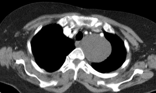
Contrast CT reveals a well-defined, solid, hypodense non-enhancing superior mediastinal mass, iso to hypoattenuating to muscle with homogeneous density, located in the left paravertebral location displacing the aortic arch branches (left subclavian, left vertebral - direct origin). Mass is indenting the oesohagus and the trachea. No vertebral erosion/ destruction.
Case Discussion
Pathologically proven ganglioneuroma.
Ganglioneuromas are fully differentiated neuronal tumors that do not contain immature elements. They tend to occur in the pediatric population and are often asymptomatic. Usually, patients are older than 10 years old, compared to neuroblastoma which occurs in patient younger than 3 years old.


 Unable to process the form. Check for errors and try again.