
Presentation
Headache and vomiting.
Patient Data
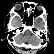

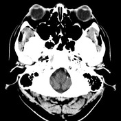
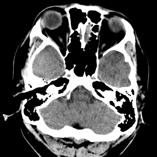
Pre and post-contrast CT brain
Findings:
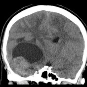
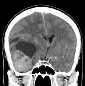
Large lesion in the right temporal lobe with a heterogeneous slightly hyperdense lobulated mass associated with a rounded well-circumscribed low attenuation component in the posterior superior aspect.
Moderate enhancement after administering IV contrast.
Associated white matter edema, although the amount of associated edema is less that what would be expected for a tumor this size.
Positive mass effect: near complete effacement of the right lateral ventricle, contralateral midline shift of 3mm with subfalcine herniation
No overlying bony permeative change.
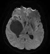
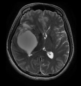

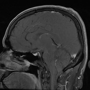

Selected MR sequences through the brain
Findings:
Confirms the large lesion in the right temporal region with solid and cystic components.
Heterogeneous enhancement of the solid component, rim enhancement of the cystic component.
Large flow void, best appreciated in sagittal projection, consistent with a large vessel traveling through the lesion
Scattered foci of enhancement in the posterior aspect of the right side of the centrum semiovale, suggestive of satellite lesions.
Fiducials are noted in both frontal and occipital regions for operative planning.
Case Discussion
This is a differential diagnosis case of a temporal lobe cystic lesion in a young adult. There are a few relevant entities one must consider when encountering a lesion of this appearance in this age group:
gangliocytoma: fully differentiated neural tumors, 60% occurring below the age of 20 years, can demonstrate solid and cystic components as well as fine, speckled calcification
ganglioglioma: although primarily occurs in a pediatric population, the incidence in young adults is higher than that of ganglioneuroma
pilocytic astrocytoma: occurring mostly in the pediatric age group, 60% are found in the cerebellum with the optic nerve, hypothalamus and brain stem consisting most of the other 40%. The location of the lesion, in this case, is somewhat unusual for a pilocytic astrocytoma
DNET: mostly occurring in the temporal lobes of young people, this is a relevant consideration, in this case
cystic metastasis: cystic metastasis with a mural nodule is the last but not least differential for this lesion
The patient went on to have surgery.
Histology
Final diagnosis: ganglioglioma (WHO grade 3) with high-grade glial component resembling glioblastoma.


 Unable to process the form. Check for errors and try again.