
Presentation
Asystole. Unsuccessful CPR attempt.
Patient Data

Endotracheal tube in oral cavity. Nasogastric tube through left nostril, appears folded in oropharynx.
Homogeneously low density of entire brain, both supra- and infratentorial, with effacement of sulci, basal cisterns, and ventricles (only thin atria of lateral ventricles visible). Consistent with diffuse, widespread brain edema due to anoxia.


SPECT with dynamic 99mTc-ECD (ethyl cysteinate dimer) injection (stack 1). During the dynamic phase, there is perfusion in the carotids bilaterally but none in the anterior and middle cerebral arteries.
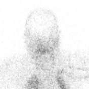
A static scan 20 minutes after 99mTc-ECD injection (stack 2) shows no radiotracer uptake (i.e. no brain perfusion) in the brain parenchyma, including the cerebral cortex, basal ganglia, cerebellum, and medulla.
The appearance of an "empty" cranium is referred to as the empty light bulb sign.
Case Discussion
Typical appearance of an anoxic, oedematic brain on CT and 99mTc-ECD (brain-specific radiotracer) SPECT scan.


 Unable to process the form. Check for errors and try again.