
Sarcoidosis
Citation, DOI, disclosures and case data
At the time the case was submitted for publication Frank Gaillard had no recorded disclosures.
View Frank Gaillard's current disclosuresPatient Data

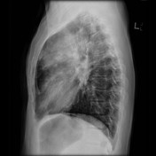
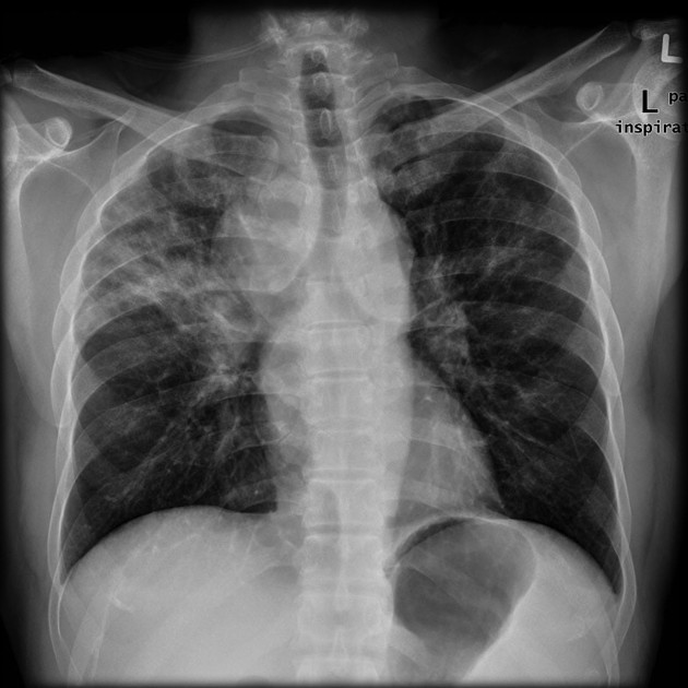
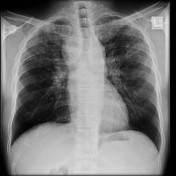
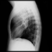
Chest x-ray demonstrates upper zone reticulonodular opacities with extensive mediastinal and hilar nodal enlargement. Stage II sarcoidosis with both nodal and parenchymal disease.

window
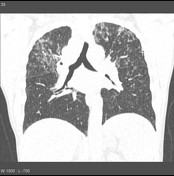
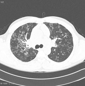
lung window
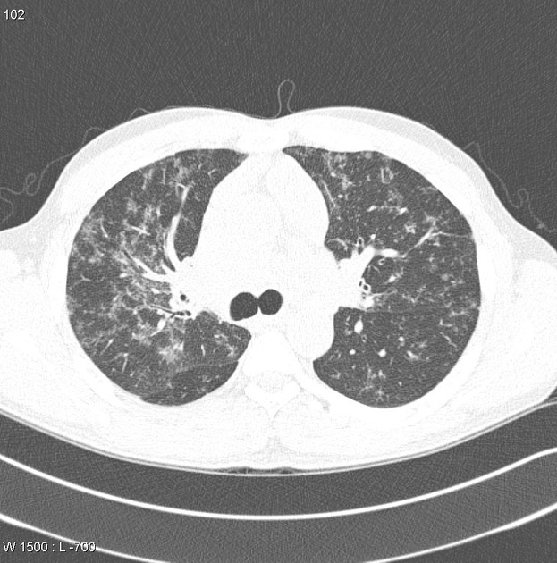
CT of the chest demonstrates diffuse areas of nodularity predominantly in a peribronchial distribution with patchy areas of consolidation particularly in the upper lobes. There is some surrounding ground glass opacities. No gross reticular changes to suggest fibrosis.
Marked mediastinal and perihilar adenopathy is present.

Xray 3 years later demonstrates some reduction on nodal enlargement.
Case Discussion
This patient had established sarcoidosis both clinically and on the basis of bronchial biopsy.
Histology
Several multinucleated giant cells identified in a chronic inflammatory, non-necrotizing background; observations are consistent with sarcoidosis.
Final diagnosis: sarcoidosis
7 articles feature images from this case
467 playlists include this case
Public playlists
- Respiratory by Pooja Kembhavi
- chest viva by saba jaradat
- Chest board cases by Savannah Shortz
- GRAFF_PLAYLISTS by Christina Dowlen
- PAH - LP Chest Viva 1 by Liam Pugh
- Exam cases by Amir Hafiz Bin Ramzah
- RAB Interstitial & Occupational by Arjuna Somanathan
- Plaman&Cardio-Vascular by Popovici Alexandra
- Diffuse Lung Disease 2024 by George Harisis
- Chest test by Gary Lee
- THX ILD/Autoimmune by Cary MacMillan
- Torax by Johana Rosinger
- Sarcoidosis by Nicholas Willmore
- HD&GL3 by Hung Dang
- Thoracic FRCR 2B by Dr Feras Salhi
- Chest_01 by Felicia Teo
- Chest by Lim Dwee Shion
- torax by Johana Rosinger
- FRCR Viva 5 by Saneej Kanhirat
- Torax by Miguel Ángel Gómez-Bermejo
- phổi kẽ by Nguyen Thi Huyen
- Spotters 2 by Abhilash Sandhyala
- Chest - interstitial by David Serich
- Chest/Vascular by eliotb
- CHEST VIVA by Juliana Tsuruta ◉
- CHEST VIVA HY by Bilal Vanlioglu
- Cleo viva 1 by Prakruthi Venkatappa
- Chest Cases by Melinda Novak
- Sarath FRCR by sarath babu
- Respiratory by Danny Chuang
- Chest 1 by Bob Ng
- v3 by Mehak un nisa parvez akhtar raja
- chest 1 by Cheng Lin Ting
- Viva Practice 2 (CPSP) by Afaaq Rasool
- resp 1 by louise lee
- Chest RANZCR by Ala Alsherbini
- a FRCR 2B viva-longs by Muthu Magesh
- Viva candidate 6 by Kavya Moonjelil Karthikeyan
- 12/10 by Fatima Abdelmoniem Abdelrahman Ragab
- UQ practice cases: Chest 5 by Craig Hacking ◉
- MD1 Respiratory by Caitlin Sweeting
- CHEST_ILD_SARCOID by Nathalie Falkner
- Chest X ray exam by Dilina Rajapakse
- Chest by Kary Suen ◈
- Viva JP by Tamer Ghorab
- Chest by Piyathida
- Revision - Chest by Aman Sandhu
- LOFTUS by Peter Playford Eriksen
- FRCR MG by Mahmoud Ghanem
- Diffuse cystic lung diseases by Dilini
- Revise by Mohamed Faragalla
- chest2 by Elsayed Amro
- Chest/thoracic Cat 1 by Gabrielle
- Chest Viva 2 by Miguel
- FRCR 2B EXCLUSIVE by PRASHANT M GITE
- Chest by Cecil Chen
- CxR Patterns by David Learmont-Walker
- ahmed chest by Ahmed Emira
- CHEST VIVA by Juliana Tsuruta ◉
- Chest Cases by Akshay Kohli
- This could be sarcoid by Sally Ayesa ◉
- 100 RX for R1 by Juan José Maya González
- chest by Muhammad Qasim Khan
- chest by SANDEEP KUMAR
- chest by Manish Chug
- Super fun mix for February by Gabrielle
- Chest by Nicholas Chen
- UIP Conditions by Timothy Jeffery
- cool simple cases by Omar Obaidat
- chest by Taqarrob abu jafar
- Chest Priya by Ayomikun Asonibare
- Chest 2b by misha kathirgamanathan
- Chest FCPS 2 VIVA by Khush Bakht
- Chest X-ray patterns by ZUL KHAIRUL AZWADI BIN ISMAIL
- Chest by Naseera Khanum
- Med Student X-ray Playlist by Inderbir Jassel
- YJL 2B Chest by YJ Lee
- Diagnostic Imaging III-Chest 1 by Sandra Norton
- Plain film exam (CXR) by Andrew Panayiotou
- Viva session 1 by Smita Patil
- TAN by Aditi Vipin
- misc chest 3 by Kevin Sheng
- Report writing practise 1 by Christine
- Facharztprüfung by Michael Jacob
- RS by Marko Atanasković
- 2B Chest by Mohammad Al-Tibi
- Chest by Ashley Hook
- M chest by Maria Javed
- sarcoidosis by Ali Labeeb Alwan
- Nodules by Nepean Radiology
- Interstitial lung disease by Ronald Joseph ◉
- Chest exam kist by Rajesh Pompapathy
- chest viva by ali alahmadi
- long cases by Fahad Alabdulghani
- Radiology teaching (Anaesthetics) by Son Do
- Aunt minnies by George Harisis
- Prüfung thorax by Lisa Ruby
- Chest tute by Rajesh Pompapathy
- Favourite chest cases by Rohit Sharma ◉
- Outras doenças pulmonares by Cleverson Leitão
- MSK by Ala Alsherbini
- Chest vivas by Sindhura Nirmalarajan ◉
- Alf VIVA Chest 1 by Kateryna Burlak
- BPT Chest by Wayland Wang ◉ ◈
- 2-Toraks by Abdullah Sukun
- diffus reticulonodular opacity by Anastasia Tjan
- 2B Chest by Mohammad Al-Tibi
- Case list 2 by Isanka Udayangani
- Chest by Piyathida
- 3. Stex Fälle A.S. by Anina Schafnitzel
- chest by Prabhat karki
- Critical Care Chest Radiology by Rebecca Dooley
- sarcoidoza by Fuiorea Iulia
- Torax by Cristian Figueroa
- mediastinum by Hamze Shaty
- Pulmonary Diseases by Ahmad Khedr ◉
- CHEST XRAY by Jyothish
- Chest by Mai-Linh Le
- Internal by Mahmoud Abusirrees
- CHEST VIVA by Juliana Tsuruta ◉
- torax by Johana Rosinger
- CHEST101 by Sherif Maccar ◉
- nezar chest by nezar shlaka
- Interstitial lung disease by Ronald Joseph ◉
- Chest exam fodder by Gabrielle
- PAH RQ 8 by Rachele Quested
- Suitable for Respiratory SCE (UK): ILD, imaging by K Ward
- Casos tórax 04/08/17 by Victor Cartaxo
- Chest reticular opacities by Xien Liversidge ◉
- Frcr medley by Sook Yin Chua
- Leicester FY1 Teaching by Kushal Joshi
- Chest by Jennifer Jones
- Chest by Joshua Yap ◉ ◈
- Chest 2B by kesy nayagam
- torax by Johana Rosinger
- Thorax by Georgia Yeo
- 100 cases 2B 30 to 50 by Dilina Rajapakse
- Chest / Vasc - 1 by James Bender
- xrays by Aoife Lee
- MAK Chest by Michael Kreltszheim
- Chest by Yue Li
- JCR 2B Chest by Jordan Curl-Roper
- viva by Heba
- WDHB - Chest by Pieter Wood
- hilar mass by nour
- Viva 4 by Marios Zertalis
- Chest by Gajan Surendra
- Charul FRCR chest practice set 4Dec by Charul Goyal
- CPSP Viva - Chest & CVS by Afaaq Rasool
- Plain films by R Srinivasan
- nove 2021 by D.D.Ranasinghe
- SQ 2B Set 1 by Shandana Qamar
- Chest viva Waseem by Waseem Mehmood Nizamani
- Chest 3 - Diffuse lung disease by Siobhan Lee
- MC Chest by Marianne Cossens
- Cat 1 chest conditions (CXR) - RANZCR curriculum by Daniel Nour
- AZ CXR ST1 TEACHING PLAYLIST by Asef Zahed
- Chest by Piyathida
- Chest 2 by N Seth
- 7-1-23 by deepti bachhuka
- long cases 1 by Jeneesh K
- Respiratory SCE by Victoria Thorley-Dickinson
- chest june 22 by elroy
- Chest VIVA by Naridchaya Aberdour
- Chest 2b by misha kathirgamanathan
- lear by Mohamad reza tarkhorani
- SEB AA Chest 1 by Sean Barrett
- Parenchymal Lung JK by Jan Kaczmarek
- FRCR 2b by Dr Priyanka Prasad R
- Viva 8 by MANIMEKALA THAMBITHURAI
- exam by 33 doctor
- ZD CVS VIVA by Dicksun Leow
- 2B Chest (order of notes) by Daoud akhtar
- Chest by Cara Lucas
- Chest by Kary Suen ◈
- CHEST (FCPS 2 viva playlist) by Kanwal Laique
- Polmone by Luca Merola
- chest by Hazal Karli
- TGRS 3 by NKOSIYAZI HLABANO
- Chest by Scott Mitchell
- Long cases by Ian Graham
- 2B: Chest by Daoud akhtar
- Edinburgh - VIVA 3 by Daniel McKernan
- 06-CHEST CASES by Rumana Hitawala
- Chest review by Ralph Nelson
- Kandy 2 by Shalitha Bandara Samarakoon
- ChestRadicools1 by Jarrel Seah
- thorax by Ioana Hutuca
- 2B CXR by GARETH MONTGOMERY
- AK FRCR 2b 2 by Amar Nitin Kanani
- 2B by Iacopo Chiavacci
- 2b chest 1 MED by R A A
- Chest Radiographs by Bilal Alam
- chest viva 1-2024 by alaa mohamed reda
- chest by Aya Aboelkhair
- Röntgen lungu by Andrea Njálsdóttir
- RESPIRATORY AND PAED PANNEL SURA by Marius Don Suranjan
- CHEST by wael
- 18/04 by RAYYA WAEL MUSA NAFFA'
- Never give up chest 1 by Fatima Abdelmoniem Abdelrahman Ragab
- Doenças Pulmonares Granulomatosas by Mariana Oliveira
- Gen Med #1 by Jen Shoobridge
- FAP 24 Thorax by Dr. Valentin Karl Ladenhauf
- CHEST FRCR by Noha Aboelenin
- CHEST FRCR by Noha Aboelenin
- Imaging 4 - Chest & Abdomen by John Bassano
- Radio 2 exam by edioardo bonamano
- Frcr 2b by Nidhi Aggarwal
- Plate 16-1-24 by Imraan Hassim
- VIVA 4 Jan 2024 by Daniel McKernan
- chest by mohammad arjmand
- Test by Sara Hanisch-Seif
- interstitial lung disease by Iulian Dragusin
- RASHED 2B CHEST by Mahmoud Rashed
- ZD Rapids Review - Thorax by Dicksun Leow
- Interstitial lung diseases 1 by marwa
- chest by Tamer
- Marwa Azab triple M mahmoud rezk by marwa
- ID Jan21 by Ashlee Kates-Ascioti
- Day 3 by Isanka Udayangani
- Chest cases by Malika Dhananjani Kumari
- 15.1 Ct Thorax Common pathology by Sahil Gulabkhan Malek
- mixed may by Jeneesh K
- mixed may by Jeneesh K
- Mediastinal Widening Differential by Nikola Grubor
- Chest cases for viva practice 25 by Snehil Kumar
- Sarcoidosis by Constantin Catanescu
- Sarcoidosis by Constantin Catanescu
- Diseases chest by marwa
- CHEST by Lucia Spoto
- CHEST by Hatem Al Jaafari
- Pneumo by TUDOR SAFIRESCU
- 01.11.24 by Gregor Peter
- Thoracic radiology by Jayathra Liyana Gamage
- cardiothoracic by reenagupta
- y decem by Shehu Mustapha
- 9 SEPT LIST2 by mehvish
- Resp Spotfires by Steven Clare
- CHEST by Hatem Al Jaafari
- FRCR 2B VIVA by Callum Watson
- marwa azab new course by marwa
- FRCR 2B VIVA by Callum Watson
- Mediastinal masses by shamini maniam
- Chest 2B by Muhammad Kaleem
- sarkoidoza by Ania Paśnikowska
- Review (Mardis) by Thornton Mardis
- CHEST VIVA 1 by Juliana Tsuruta ◉
- chest viva cases by Abdulrahman abdo ali abbas
- chest viva cases by Abdulrahman abdo ali abbas
- Marwa Azab long cases examples by marwa
- FA lunge by Zayn Dh
- CXR - lungs by Hari Prasath Sivakumar
- Interstitial lung disease by Reham Mohamed
- Marwa Azab Approach chest by marwa
- Prüfung3 by Marie Hölting
- A 2021 long by Abdullah Hajar
- thorax by Gevorg Kurkdzian
- 24.2/25 by Hari Prasath Sivakumar
Unlisted playlists
This case is used in 209 unlisted playlists.
Related Radiopaedia articles
Promoted articles (advertising)
How to use cases
You can use Radiopaedia cases in a variety of ways to help you learn and teach.
- Add cases to playlists
- Share cases with the diagnosis hidden
- Use images in presentations
- Use them in multiple choice question
Creating your own cases is easy.
ADVERTISEMENT: Supporters see fewer/no ads


 Unable to process the form. Check for errors and try again.