
Presentation
Suspicion of CPAM on prenatal ultrasound.
Patient Data

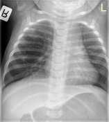
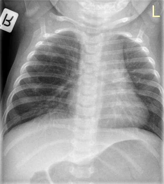
Chest X-ray taken on the day of birth.
Central opacities. Right perihilar lucencies, more prominent (pseudocystic) in the upper zone. The differential diagnosis includes CPAM.
No pleural effusion.
Cardiac silhouette normal, small thymus.
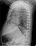
Chest X-ray taken at age 6 months.
Large cystic lesion in the RLL.
Left lung well aerated.
Cardiac silhouette of normal size.
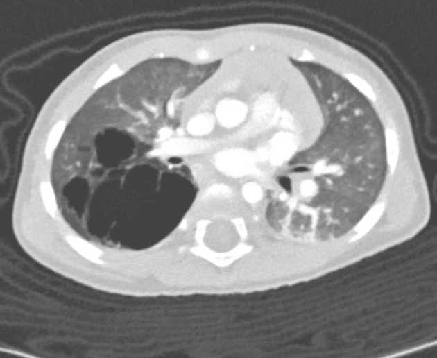
The upper segment of the RLL is very large and is composed of cysts of different sizes, ranging up to a diameter of 2.5 cm. The entire lesion measures 4.8 x 4.1 x 4.7 cm. Mild adjacent pleural thickening. No soft tissue component or abnormal vasculature. Small RUL atelectasis. Small atelectases in the dependent portions. Mild tracheal compression anteriorly by the brachiocephalic artery.
In summary: features consistent with CPAM.
Case Discussion
Pathology report:
Upper segment right lower lobe:
Features compatible with congenital cystic adenomatoid malformation, type I.


 Unable to process the form. Check for errors and try again.