
Presentation
Presented with a gradual loss of extension and abduction for the right thumb during the last 2 to 3 weeks. Associated with the right proximal forearm pain. No significant trauma. Referred to check the extensor pollicis longus tendon injury.
Patient Data
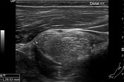


There is a well-defined, encapsulated lesion at the anterior radial side of the proximal forearm. It is hyperechoic in comparison to the adjacent muscle along with the presence of linear internal echogenic strands. There is no calcification/ cystic changes/ vascularity. The approximate size of the lesion is about 31 x 29 x 15 mm. With compression, the lesion is deformable.
The lesion is between the superficial and deep heads of the supinator muscle causing anterior displacement and compression of the posterior interosseous nerve resulting in nerve edema and hypoechogenicity.
The asymptomatic side posterior interosseous nerve was examined for comparison which shows normal coarse, normal echopattern without any perineural mass lesion.
Extensor pollicis longus tendon is intact. There are possibly two tendon slips just distal to Lister's tubercle. The extensor muscles show moderate diffuse fatty infiltration and moderate volume loss as compared to the asymptomatic side.
The common extensor tendon shows normal echopattern without changes of tendinosis or tear.
Case Discussion
A young male developed right thumb extension and abduction weakness during the last few days. Clinically, extensor policies longus tendon pathology was suspected.
Ultrasound shows a normal echopattern of the extensor pollicis longus tendon. There is a lipoma between two heads of the supinator muscle resulting in posterior interosseous neuropathy. There are secondary changes in the innervated muscles. Lateral epicondylitis is a differential diagnosis in such a case. The ultrasound shows a normal echopattern of the common extensor tendon.
There is no surgical follow up of this case.
Ultrasound is now considered as the first-line modality for the evaluation of the peripheral nerves of the upper extremity. Ultrasound has many advantages over magnetic resonance imaging like higher soft-tissue resolution, low cost, dynamic imaging, a quick examination of a whole limb, comparison with the asymptomatic side1.
Check out this companion case (rID 63343) with surgical follow-up.


 Unable to process the form. Check for errors and try again.