
Mitochondrial encephalomyopathy, lactic acidosis, and stroke-like episode (MELAS)
Presentation
Seizures and lactic acidosis.
Patient Data
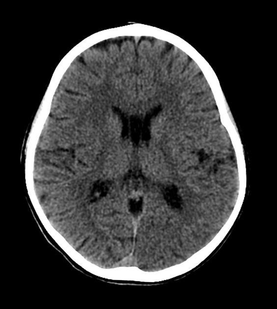
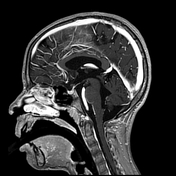
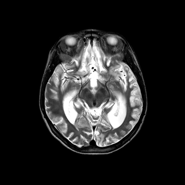
Selected images demonstrate a small area of hypoattenuation involving the cortex and subcortical white matter in the left temporal lobe.


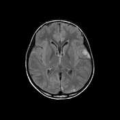


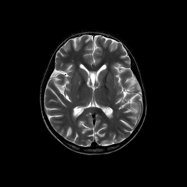
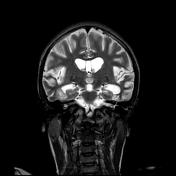
Selected MRI images demonstrating confluent areas of T2/FLAIR hyperintense signal of the left temporal and occipital cortex with increased signal on DWI, corresponding to little change on ADC. In addition, moderate gyriform enhancement.
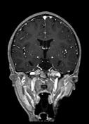
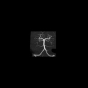
On MRA images, normal appearance of the intracranial vessels, with no signs of vasculitis.
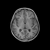
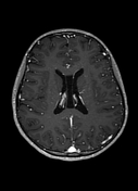
MRI, about 1.5 years letar

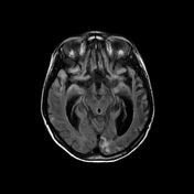
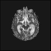
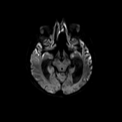
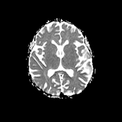
Selected images demonstrating confluent areas of gliosis throughout the cerebral hemispheres in keeping with sequelae of previous ischemic insults. In addition, advanced atrophic changes with ex-vacuo dilatation of the ventricles.
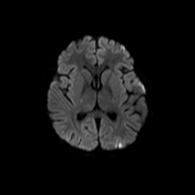
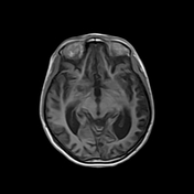
Evidence of restricted diffusion at several cortical regions, in particular in the insular/perisylvian and frontal regions, in keeping with ongoing ischemic insults.
Case Discussion
Recurrent episodes of seizure, unexplained decreased level of consciousness and associated with lactic acidosis, overall suggestive of mitochondrial disease. MELAS was confirmed by genetic testing.
MELAS is characterized by reoccurring stroke-resembling events, in non-vascular territories, which are assumed to be secondary to mitochondrial dysfunction. After several events, we noticed a rapidly progressing brain atrophy.
Our patient also had diabetic mellitus, which can be well explained by mitochondrial dysfunction.
The possibility of MELAS should be addressed in the differential diagnosis of acute strokes in young patients, especially if the pattern doesn’t follow the typical vascular territories and/or the vascular imaging is unremarkable.
With courtesy of Dr. Christine Saint-Martin, Associate Professor in Neuroradiology and Pediatric Radiology at McGill University.


 Unable to process the form. Check for errors and try again.