Presentation
Pain around knee.
Patient Data

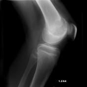
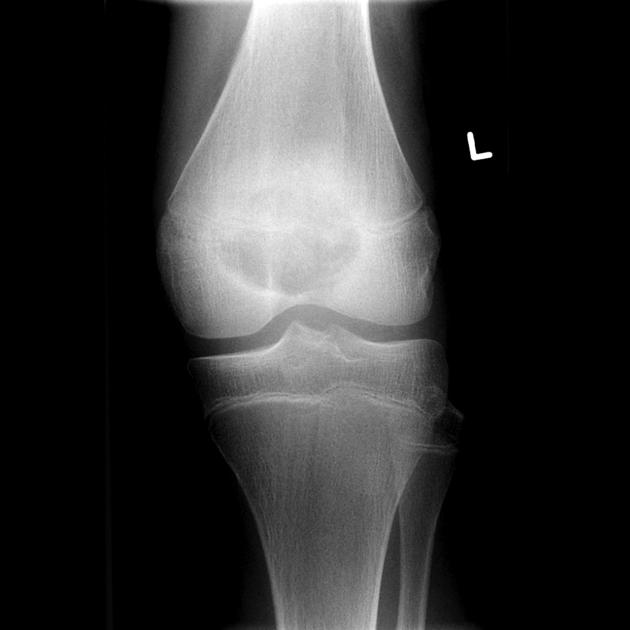
A sharply defined lucent lesion is centered on the epiphysis of the distal femur, and appears to transgress the growth plate (which remains open). It has a narrow zone of transition and no convincing matrix calcification. Anteriorly it appears to abut the articular surface, possibly breaching it. A joint effusion is present. No periosteal reaction is present.

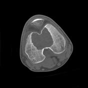
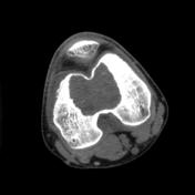
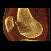
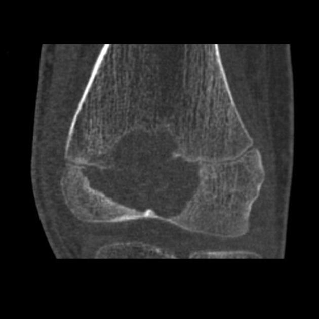
CT confirms the plain film appearances, revealing a sharply demarcated epiphyseal lucent lesion but with faintly sclerotic margins. It transgresses the growth plate into the anterior part of the metaphysis. There is no periosteal reaction, however the does appear to be a cortical breach anterosuperiorly into the knee joint. No matrix calcification or extra-osseous soft tissue component can be demonstrated.
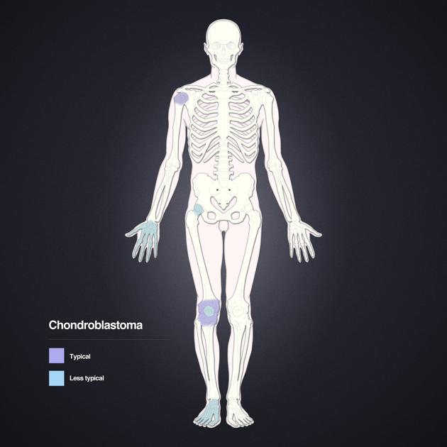
Distribution of chondroblastomas. Layout and distribution: Frank Gaillard 2012, Line drawing of skeleton: Patrick Lynch 2006, Creative Common NC-SA-BY
Case Discussion
This case demonstrates typical appearances of a chondroblastoma, in a typical location. The lesion was curetted and pathology confirmed the diagnosis.


 Unable to process the form. Check for errors and try again.