
Presentation
Three month history of spinal pain, worse in the thoracic spine. Pain worsens with rest and improves with activity. Raised ESR 35. No recent history of trauma. CXR to rule out any infective cause.
Patient Data
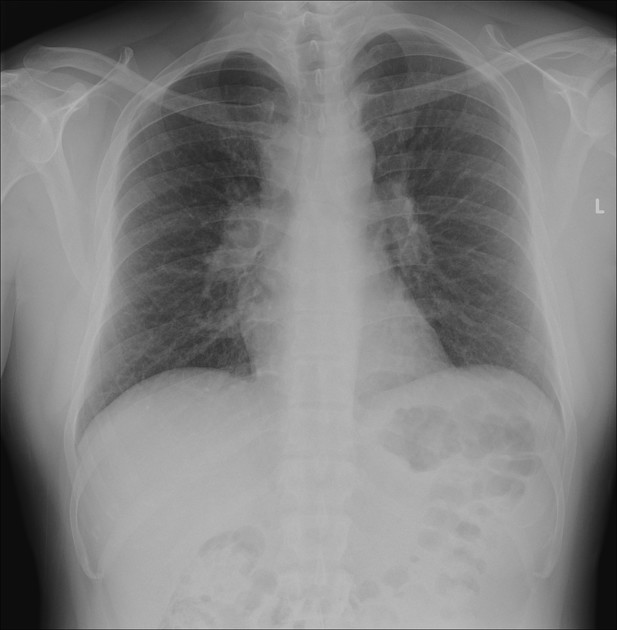
Garland triad of bilateral hilar lymphadenopathy and right paratracheal lymphadenopathy.
Heart size normal. Lungs clear.
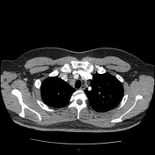

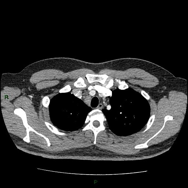
Extensive mediastinal lymphadenopathy at all nodal stations, with conglomerate lymph nodes in the para/pretracheal space. The largest discrete node is at the right hilum measuring 5 cm. Multiple peribronchial nodes, the largest 1.3 cm in the right lower lobe.
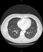
Two 3 mm nodules in the left lower lobe, one of which is subpleural. No perifissural nodularity or fibrosis.
No supraclavicular, axillary or cervical lymphadenopathy.
The solid organs are normal. No infra diaphragmatic lymphadenopathy. No bony or paraspinal soft tissue abnormality.
Case Discussion
The conclusion from CT imaging: extensive mediastinal lymphadenopathy – I suspect this is due to sarcoidosis.
If a tissue diagnosis is required, the nodes are amenable to endobronchial ultrasound (EBUS) biopsy.
EBUS procedure and histology:
- mediastinal node FNA: AFFB negative
- EBUS of mediastinal node: non-caseating granulomatous inflammation
Final diagnosis: sarcoidosis.


 Unable to process the form. Check for errors and try again.