
Presentation
Progressive dysphagia with chest pain. Rule out malignancy.
Patient Data
Age: 35 years
Gender: Female
From the case:
Achalasia


Download
Info
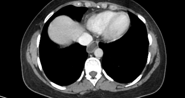
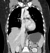
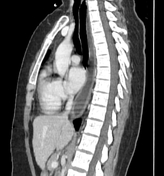
Grossly dilated esophagus with a thin and regular wall, filled with fluid/food debris with bird beak sign at the gastro-esophageal junction.
No mediastinal or hilar lymphadenopathy is seen. Both lungs are clear (lung window not shown).
Case Discussion
CT appearance of an achalasia.
CT has little role in directly assessing patients with achalasia, but is useful in the assessment of the esophageal wall to identify any focal thickening which may indicate malignancy.


 Unable to process the form. Check for errors and try again.