
Presentation
Obstructive jaundice, epigastric pain and tenderness.
Patient Data
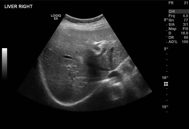
The pancreas shows heterogeneous echopattern with suggestion of two masses measuring 2.3 cm and 2 cm.
Dilated CBD measuring 1 cm in caliber and intra-hepatic biliary radicals dilatation is also noted.
The gallbladder has sludge and shows diffuse wall thickening. No stones are seen.
The rest of the examination is unremarkable. (images not included)
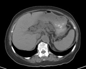


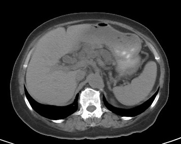
The pancreas is enlarged and contains numerous different sized cystic lesions, the largest measures 2.8 x 2 cm. No evidence of solid pancreatic mass lesions could be seen.
Dilated common bile duct and intrahepatic biliary radicals with no evidence of adjacent organ invasion, vascular encasement, or invasion.
The gallbladder has a thick enhancing wall with pericholecystic edema suggestive of chronic cholecystitis.
No evidence of enlarged peritoneal lymph nodes could be seen.
The liver, pancreas, adrenal glands, and kidneys appear normal.
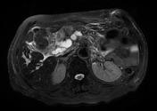
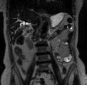
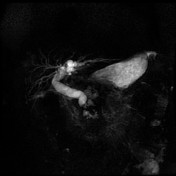

Multiple different sized cystic lesions are seen in the pancreatic head which appears not communicating with the pancreatic duct.
The common bile duct and intrahepatic bile ducts are seen grossly dilated.
The maximum transverse diameter of the CBD is 1.7 cm.
The maximum transverse diameter of the left main duct is 9.5 mm.
There is no evidence of filling defects to suggest stones.
The distal end of the CBD appears tapering and is not visualized for the last 1 cm.
The findings are suggestive of a tight stricture rather than stones.
Case Discussion
This 70 Y/O woman presents with epigastric pain, tenderness, and obstructive jaundice. Initially, an ultrasound of the abdomen was requested and showed a heterogeneous echo pattern of the pancreas with associated CBD and intrahepatic biliary radicals dilatation, further evaluation by CT, showed multiple cysts involving the pancreas, but with no evidence of solid lesion. So it was thought to represent branch duct Intraductal papillary mucinous neoplasms (IPMN). However, MRCP did not demonstrate communication with the pancreatic duct, and a biopsy was advised.
Histology:
MACROSCOPIC DESCRIPTION: Whitish tumor measuring 3 cm in maximum dimension involving the wall of the common bile duct and surrounding pancreatic tissue.
MICROSCOPIC DESCRIPTION: Sections reveal a pancreatic head ductal adenocarcinoma in the pancreatic head, composed of complex crowded infiltrative glands lined by pleomorphic columnar epithelium surrounded by desmoplastic stroma. The tumor is invading the duodenal wall reaching the submucosa and involving the base of the ampulla. The tumor is encroaching upon the intrahepatic portion of the common bile duct. Foci of perineural and lymphatic invasion are seen. All margins of resection are free.
FINAL DIAGNOSIS: Distal stomach, head of pancreas and duodenum, Whipple resection: Ductal adenocarcinoma of the pancreatic head.


 Unable to process the form. Check for errors and try again.