
Presentation
Work up for hematuria.
Patient Data
Age: 55 years
Gender: Female
From the case:
Renal cell carcinoma


Download
Info
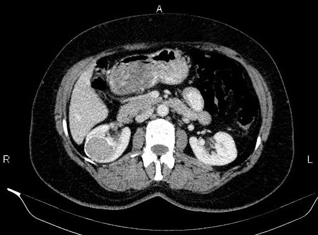
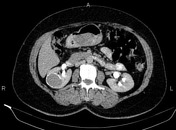
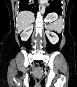
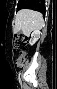
A 40×32 mm well-defined oval-shaped low enhancing mass with incomplete peripheral ring calcification is noted at mid-portion of the right kidney. There is no sign of local invasion to adjacent structures and no obvious vascular extension. Perinephric fat is intact. No regional lymphadenopathy could be seen.
A few parapelvic cysts are seen at left kidney less than 20mm.
Degenerative changes as osteophytosis are seen at the lumbar spine.
Case Discussion
Right renal mass; pathology proven renal cell carcinoma.
CT is frequently used to both diagnose and stage renal cell carcinomas. Approximately 30% demonstrate some calcification 1.


 Unable to process the form. Check for errors and try again.