
Presentation
Abdominal pain and dyspepsia.
Patient Data
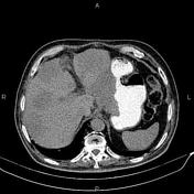

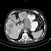
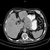
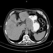
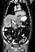
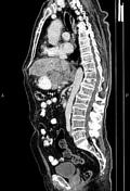

Increased wall thickness due to tumoral infiltration is present at gastric cardia and proximal of lesser curvature. Multiple lymphadenopathies are noted in the vicinity of the diseased segment as well as parailiac, peripancreatic and para-aortic regions. Additionally, multiple low enhancing masses are noted at the liver less than 65 mm.
Several small parapelvic cysts are seen at left kidney.
The prostate gland is enlarged.
Degenerative changes as osteophytes are seen at the thoracolumbar spine. Anterior wedge compression is present at T12 vertebral body.
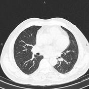
Several small nodules, mostly subpleural, are seen at both lungs less than 5 mm.
Case Discussion
Gastric mass; pathology proven adenocarcinoma with lymphadenopathies; hepatic and lung metastasis.
CT is currently the staging modality of choice because it can help identify the primary tumor, assess for the local spread, and detect nodal involvement and distant metastases.


 Unable to process the form. Check for errors and try again.