Presentation
Asymptomatic.
Patient Data
Note: This case has been tagged as "legacy" as it no longer meets image preparation and/or other case publication guidelines.

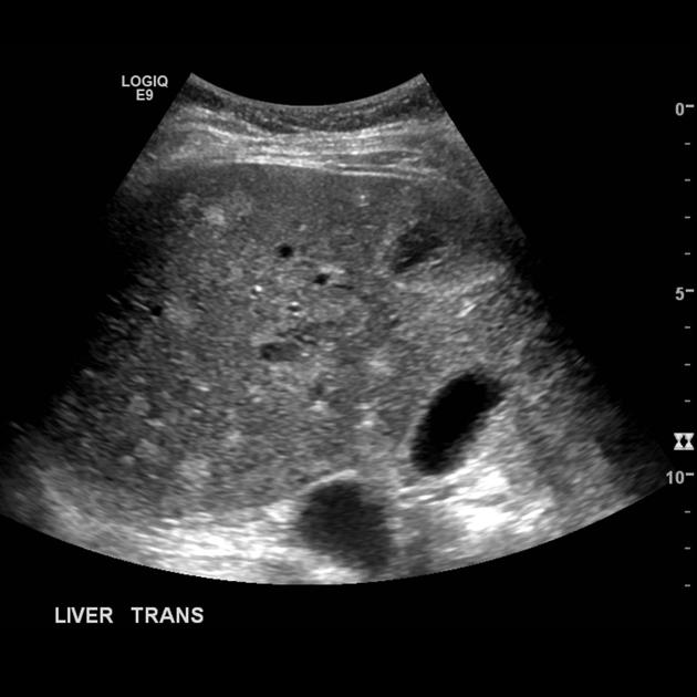
Multiple Biliary Hamartoma. Ultrasound shows many small round echogenic liver lesions and a few larger hypoechoic lesions.
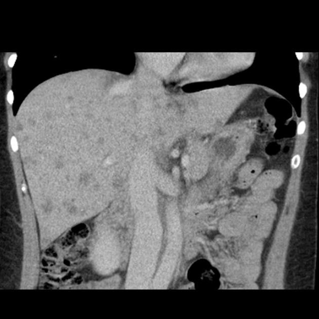
Portal venous phase CT showing multiple small hypoattenuating liver lesions.
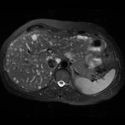
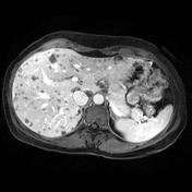
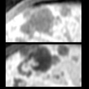
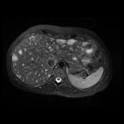

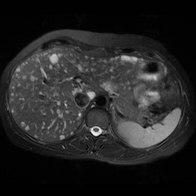
MR images show multiple small T2 hyperintense and T1 hypointense liver lesions. Several of the lesions have an enhancing periphery or enhancing nodule.
Case Discussion
Ultrasound, CT and MRI demonstrate numerous liver lesions.
On ultrasound they are generally small and echogenic with a few larger hypoechoic lesions. CT showed many small 3-25 mm hypoattenuating lesions. MR revealed the lesions to be T2 hyperintense and T1 hypointense. Most lesions did not enhance however some contained an enhancing nodule or peripheral rim of enhancement.
Features were thought to be most consistent with multiple biliary hamartomas (von Meyenburg complexes).


 Unable to process the form. Check for errors and try again.