
Presentation
Abdominal pain and dyspepsia.
Patient Data
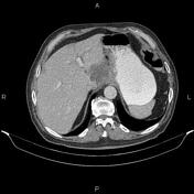

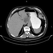

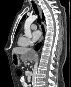

Increased wall thickness due to tumoral infiltration is present at the distal third of the esophagus. Tumoral infiltration is also observed at the esophagogastric junction and gastric cardia. Additionally, a 77×57 mm mass is present between the gastric lesser curvature and left lobe of the liver that originated from the gastric wall and invades adjacent liver parenchyma.
Regional lymphadenopathy is evident. Lymphadenopathy with SAD of 17 mm is also present in the subcarinal region, and a small one with SAD of 10 mm is noted in the upper right paratracheal region.
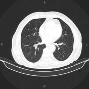
A 7 mm subpleural nodule is evident at the right middle lobe.
In addition, several small low enhancing masses, less than 15 mm, are seen in the liver, suggestive of metastasis.
A few nonenhanced simple cortical cysts are seen in both kidneys.
The prostate gland is enlarged.
Case Discussion
Esophagogastric tumoral infiltration; pathology proved adenocarcinoma with mediastinal and perigastric lymphadenopathy, local invasion to the left liver lobe, and liver and lung metastases.


 Unable to process the form. Check for errors and try again.