
Presentation
Work up for dyspepsia.
Patient Data
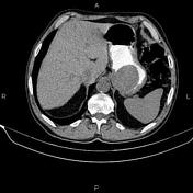

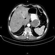

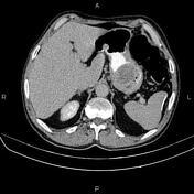
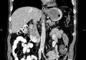
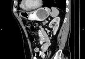
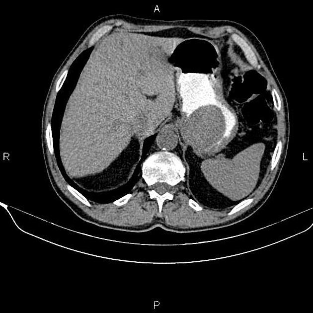
A 65×50 mm well defined intraluminal mass is present at the gastric fundus and proximal of the body. Focal defect is present at the right lateral aspect of the mass and could suggest an ulcer.
Inhomogeneously, the hepatic attenuation value is less than the spleen, suggesting uneven fatty liver.
Linear calcification is present at the lateral aspect of the spleen.
Several nonenhanced simple cortical cysts are seen in both kidneys.
Case Discussion
The patient's gastric mass resection and histopathology evaluation confirmed gastrointestinal stromal tumor (GIST) without malignant transformation.
It is difficult to differentiate a benign from a malignant GIST radiologically, but exogastric growth, diameter >5 cm, central necrosis, and extension to other organs could suggest malignant transformation.


 Unable to process the form. Check for errors and try again.