
Presentation
Patient presenting with abdominal pain, fever, tachycardia, and weight loss. Palpation of mass below the left scapula.
Patient Data


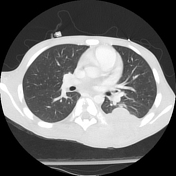
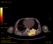
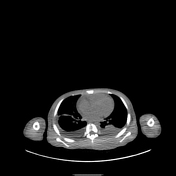
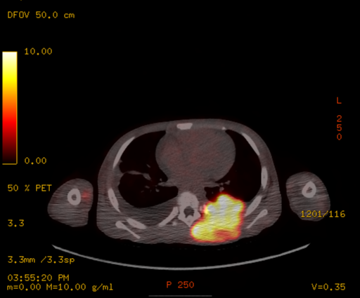
There is a large left chest wall heterogeneously enhancing mass with aggressive periosteal reaction involving primarily the left posterior seventh rib and also adjacent fifth, sixth, and eighth ribs. The mass extends into the left thoracic cavity and into the paraspinal muscles. There is no definitive
component extending into the spinal canal. There is pleural nodular thickening near the costovertebral junctions of the third and fourth ribs.
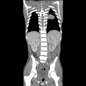
There is lymphadenopathy involving both the thorax and abdomen.
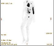
There are multiple FDG avid left neck level 3 through 5 and left supraclavicular lymph nodes are present. There are FDG avid intramuscular metastases are seen at the level of the lower cervical spine and left paraspinal metastasis at the level of C7 .
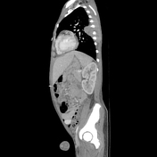
There is an intensely FDG avid posterior left chest wall mass is seen extending from the
level of T3-T10. The mass extends posteriorly to the level of left chest subcutaneous fat and anteriorly where it invades pleura and extends into the left lung lower lobe. There is intense radiotracer uptake within the posterior aspect of the left fourth through through eighth ribs.
Incidental butterfly vertebra at T6.
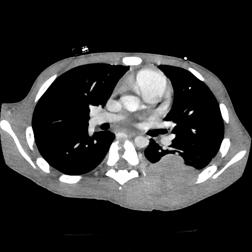
FDG avid right pleural metastases are also seen at the level of the both upper lobes as well as much more intensely at the level of the medial right lung lower lobe superior segment and superior left mediastinal, right retropectoral, and right axillary lymph nodes.
There are multiple FDG avid mesenteric, retroperitoneal, and right common iliac lymph nodes.
Case Discussion
This is a case of a T cell acute lymphocytic lymphoma. Biopsy of the mass was performed for pathologic analysis. Cytogenetic analysis revealed a NPM1-ALK fusion. A subsequent bone marrow aspirate detected a trace population of atypical cells that were positive for CD2, CD4, CD5, CD7, and CD22 (dim), and CD45.
Co-author:
Ping Lu, MD
Travis Bevington, MD


 Unable to process the form. Check for errors and try again.