
Presentation
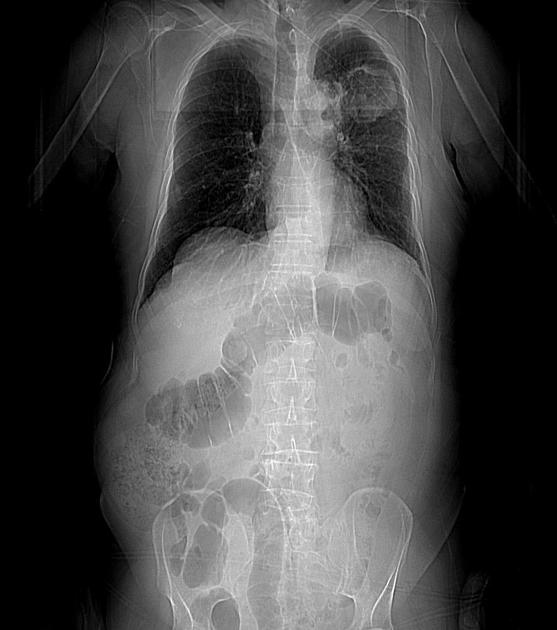
Transitional cell carcinoma of the urinary bladder referred with the new onset dyspnea.
Patient Data

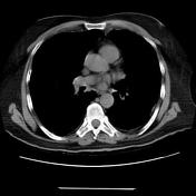


Two large cavities with thick irregular walls along with left upper lobe apicoposterior and anterior segments associated with adjacent irregular thickening of interlobular septae.
Close contact of cavities with aortic arch but the fat plane is preserved.
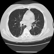
Reticular bands and fine subpleural fibrotic changes along with both lower lobes superior and posterobasal segments.
Air cysts within the right lung parenchyma.
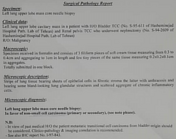
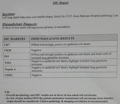

Microscopic diagnosis was compatible with non-small cell carcinoma (primary or secondary).
Case Discussion
Regarding the above imaging and pathological findings, metastatic transitional cell carcinoma to the lung is the most probable diagnosis.
Original case courtesy of Dr. Behrooz Emami Mehr.


 Unable to process the form. Check for errors and try again.