Presentation
Known case of diabetes mellitus, hypertension and benign prostatic hyperplasia (BPH).
Patient Data

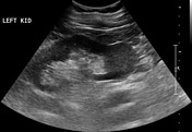
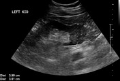
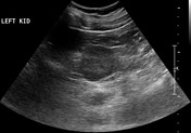
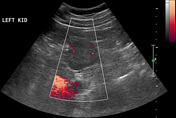
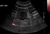
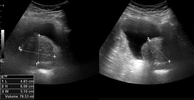
Enlarged heterogeneous prostate gland (BPH) measures 5.3 x 5.2 x 5.3 cm with an average volume of 75 ml. Incidental finding of a well-defined, isoechoic, rounded mass lesion measuring approximately 3.6 x 4.0 cm at the lower pole of left kidney. Some internal vascularity is appreciable in it on color Doppler ultrasound examination.
Findings: An enhancing exophytic mass lesion measuring about 3.7 x 4.7 cm is seen at the lower pole of the left kidney. A few small necrotic areas are seen in it. Patent renal vessels and IVC. No evidence of metastases is noted. A few simple bilateral renal cortical cysts. Fat stranding & subcentimeter lymph nodes around the mesenteric vessels, suggestive of mesenteric panniculitis. A calcified nodule measuring 10 mm is seen in the left upper lobe which is likely an old pulmonary granuloma. Bilateral pars interarticularis fracture (spondylolysis) at L5 level with grade I spondylolisthesis at L5/S1 level. Enlarged prostate gland. Impression: Enhancing exophytic left renal mass lesion suspicious of renal cell carcinoma.
Case Discussion
Pre-operative diagnosis: Left renal mass.
Procedure: Laparoscopic left radical nephrectomy.
Diagnosis: Histologic type: Clear cell renal cell carcinoma. Tumor size: Tumor size: 4 x 3.6 x 1.5 cm. Histologic grade: grade 1 (Fuhrman nuclear grade). Tumor focality: Unifocal. Macroscopic extent of tumor: Tumor limited to kidney. Sarcomatoid features: Not identified. Tumor necrosis: Present. Microscopic tumor extension: Tumor limited to kidney. Margins: Uninvolved by invasive carcinoma. Lymphovascular invasion: Not identified.
Pathologic staging: pT1a, pNx, pMx


 Unable to process the form. Check for errors and try again.