
Presentation
Iron deficiency anemia. Endoscopy suspicious for gastric carcinoma. Gastric outlet obstruction?
Patient Data


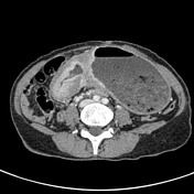

Long circumferentially thickening segment of antrum and pylorus with a wall thickness exceeding 1 cm.
The stomach is markedly distended containing food residue.
Perigastric fat stranding and small volume lymphadenopathy up to 7 mm at the site of the thickened gastric wall.
The solid organs are normal.
Multiple calcified gallstones within the gallbladder.
Case Discussion
The appearances are typical of a gastric adenocarcinoma involving the distal stomach resulting in gastric outlet obstruction.
Typical modes of presentation are iron deficiency anemia (as in this case), weight loss or vomiting.
The stomach wall is much better appreciated with a distended stomach lumen (ideally water as a negative contrast agent) when assessing it with CT.
ENDOSCOPIC BIOPSY: Diffuse infiltrating gastric adenocarcinoma


 Unable to process the form. Check for errors and try again.