
Presentation
Chronic constipation, acute lower abdominal pain, leukocytosis? diverticulitis.
Patient Data
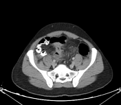

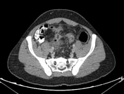
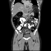
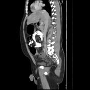

Segmental stenotic lesion of the sigmoid colon with significant luminal compromise and subsequent proximal colonic obstruction with competent ileocecal valve.
Marked fat, edema, with abscess adjacent to the diseased segment of the sigmoid inferring secondary inflammatory changes likely on top of focal wall perforation.
Multiple paracolic and mesenteric small lymph nodes.
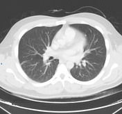
Multiple hepatic focal lesions and pulmonary nodules.
Case Discussion
Paracolic inflammation and abscess prompted an investigation for an underlying lesion, particularly in the setting of colonic obstruction. A stenotic lesion in the sigmoid colon wall was identified, highly suggestive of malignancy.
Although rare, this presentation is documented in the literature as an uncommon manifestation of colonic malignancy. It typically results from transmural invasion of the colonic wall and elevated intraluminal pressure, leading to perforations. In this case, the presence of colonic obstruction, along with hepatic and lung metastases, strongly suggested malignancy. Otherwise, the CT picture can closely mimic diverticulitis.
The patient was transferred to a specialized oncologic center for further evaluation and management. No additional data were available at the time of reporting.


 Unable to process the form. Check for errors and try again.