
Presentation
Posterior parietal swelling in a female patient with breast cancer.
Patient Data


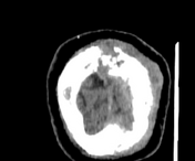
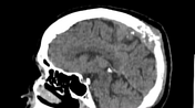
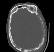
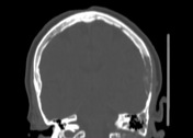
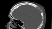
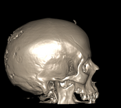
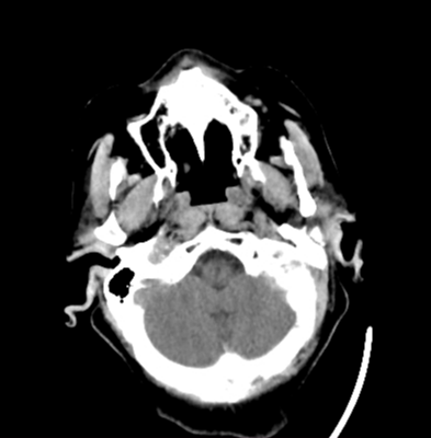
Corresponding to the site of clinical complaint at the mid-posterior parietal region, there is an osteolytic destructive bony lesion associated with extraosseous intra & extracranial soft tissue components. Other mixed lytic and sclerotic bony lesions are scattered in the skull bones.
Two other incidental findings are noted:
symmetrical thickening of the inner skull vault at both frontal regions in keeping with hyperostosis frontalis interna
a small well-defined partially calcified scalp mass at the mid-frontal region in keeping with proliferating trichilemmal cyst
No intracerebral SOLs can be detected.
Case Discussion
Considering the patient history of breast cancer, bony lesions were diagnosed as mixed lytic and sclerotic bone metastasis, which is one of the patterns of bone metastasis commonly seen in breast cancer.
It can also be seen in other primary malignancies including lung carcinoma, carcinoma of the cervix, testicular tumors, prostate carcinoma, ganglioneuroblastoma, gastrointestinal cancers, and squamous cell carcinomas.


 Unable to process the form. Check for errors and try again.