
Presentation
Cough.
Patient Data
Age: 65 years
Gender: Male
From the case:
Solitary pulmonary nodule - lung cancer
Show annotations
Download
Info
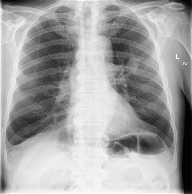
3 cm left upper lobe mass in the perihilar region.
Heart size normal. Right lung clear. Normal mediastinal contours.
Case Discussion
The typical appearance of a solitary pulmonary nodule with some practical tips:
there is a differential, but it is lung cancer until proven otherwise
at 3 cm in size, this is a "mass", not a "nodule" (which measures < 3 cm)
be sure to check there is only one nodule
ensure there are no other clues, such as an AP window node
always check prior imaging - unnecessary anxiety and cost through CT scanning can be avoided if the mass has been there for years and has a benign cause, such as a hamartoma


 Unable to process the form. Check for errors and try again.