
Presentation
Pain left hemiscrotum. No urinary symptoms.
Patient Data
Age: 35 years
Gender: Male
From the case:
Testicular seminoma

Download
Info
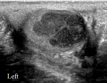
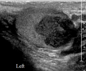
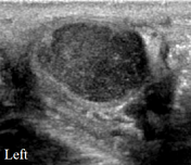
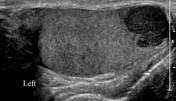
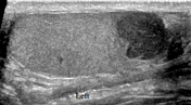
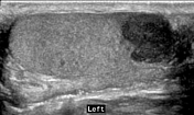
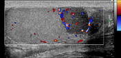
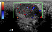
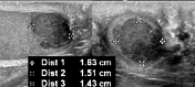
A well-defined lobulated hypoechoic eccentric nodule measuring 1.6 x 1.5 x 1.4 cm, is seen in the left testis. No cystic component or calcifications are seen in it. Color Doppler ultrasound examination shows internal vascularity.
Download
Info
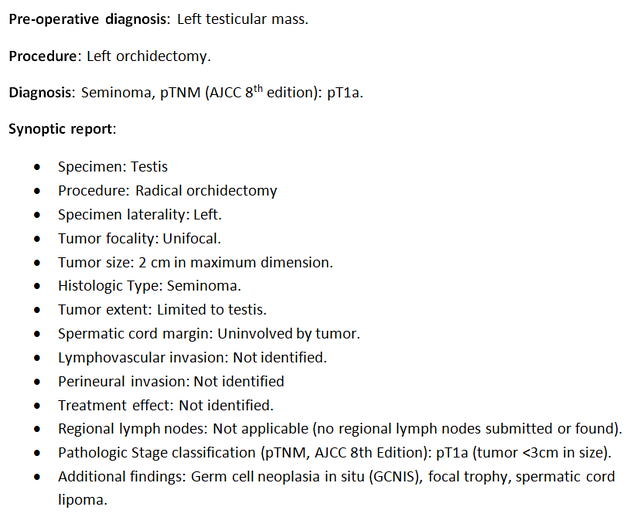
Histopathology of the left orchiectomy showing 2 cm seminoma.


 Unable to process the form. Check for errors and try again.