
Presentation
Known case of Tetralogy of Fallot. Status-post palliative shunt surgery.
Patient Data

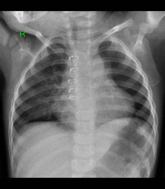
There is unequal pulmonary vascular markings with the right lung exhibiting increased vascularity suggestive of preferential blood flow and the left lung with decreased vascularity.
Heart shows uplifted apex reflective of right ventricular hypertrophy.
Sternotomy wires from prior surgery are seen.
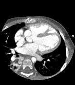

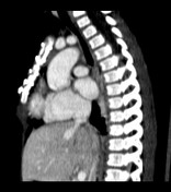
The heart shows right ventricular hypertrophy.
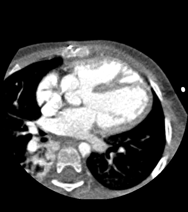
Right ventricular outflow tract obstruction is demonstrated in the form of infundibular stenosis.
There is a ventricular septal defect and overriding dilated aortic root.
Both the right and left main coronary arteries arise from the anterior and posterior coronary sinus, respectively. The non-coronary sinus is at the right lateral aspect of the root. The left divides into the anterior descending and circumflex arteries. No evidence of coronary artery stenosis or aneurysmal dilatation is identified.
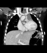
The post-surgical palliative shunt connecting the innominate and main pulmonary artery is patent.
There is moderate to severe stenosis at the proximal left pulmonary artery resulting to preferential blood flow into the right lung.
All pulmonary veins drain into the left atrium.
The inferior vena cava and a single superior vena cava drain into the right atrium.
Serpiginous vessels are identified in the pericarinal region and around the proximal bronchi probably from collateral vessel formation.
There are areas of atelectasis in the right lung.
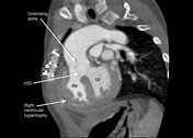
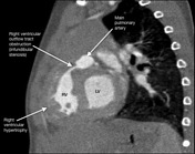
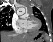

Annotated CT images demonstrating the components of tetralogy of Fallot (TOF):
- right ventricular hypertrophy
- right ventricular outflow tract obstruction (RVOTO) in the form of infundibular stenosis
- ventricular septal defect (VSD)
- overriding aorta
A modified Blalock-Thomas-Taussig shunt is also shown connecting the innominate artery and main pulmonary artery.
Case Discussion
Tetralogy of Fallot (TOF) is a congenital cardiovascular anomaly that consists of right ventricular outflow tract obstruction, right ventricular hypertrophy, high ventricular septal defect (VSD) and an overriding aorta. Hemodynamically, the critical component of the anomaly is the right ventricular outflow tract obstruction which may come in a range of presentations from mild pulmonary stenosis to pulmonary atresia. In the past, the patient in this case underwent palliative shunt surgery (modified Blalock-Thomas-Taussig shunt) to improve pulmonary circulation and to prepare the pulmonary arteries for total corrective surgery. However, peripheral stenosis is present at the proximal left pulmonary artery which resulted to unequal blood flow (preferential flow to the right lung and decreased flow into the left lung) which is evident in the accompanying chest radiograph.


 Unable to process the form. Check for errors and try again.