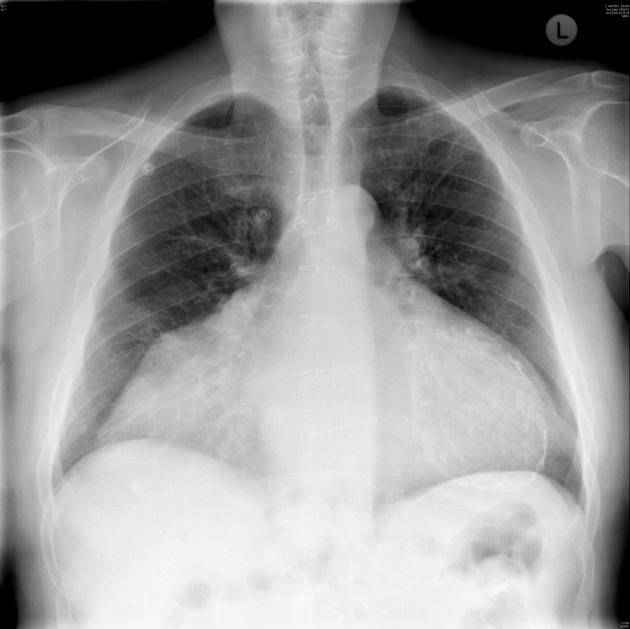
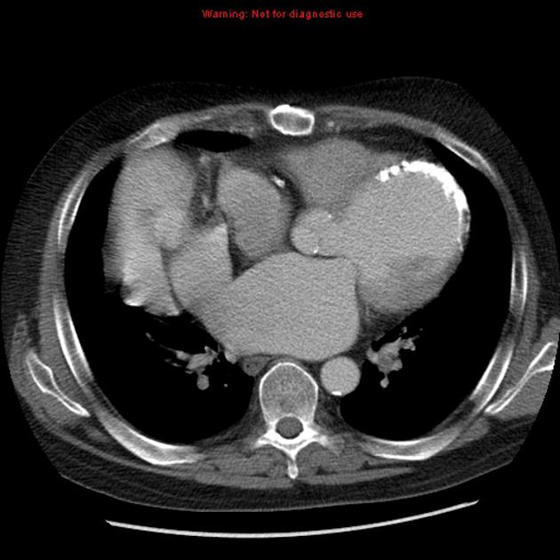
Heart transplantation is a major surgical procedure for acute or chronic end-stage heart disease in which the failing heart is replaced with a healthier donor heart. It requires a comprehensive multidisciplinary approach and radiology plays a major role in the evaluation of patients before and after heart transplantation.
This article does not discuss the complex medical issues around heart transplantation such as immune typing and post-operative immunosuppression.
On this page:
Indications
There are several indications for transplantation and many require the exhaustion of non-surgical alternatives before transplantation is considered:
-
the majority of adult transplants are for cardiomyopathy 1
non-ischemic etiologies are a more common indication than ischemia
valvular disease and cardiomyopathy
infiltrative myocardial disease e.g. sarcoidosis
cardiac tumors
congenital heart disease is the most common indication in pediatric patients 1
Types of heart transplantation
There are two main types of heart transplantation performed with medial sternotomy 1:
-
orthotopic heart transplant - the deceased donor heart is transplanted into the recipient's pericardial sac immediately following removal of the diseased heart from the recipient. This is the most common type and there are two basic methods:
biatrial method - the recipient’s heart is removed with a portion of the native atria (atrial cuff) left in situ to be anastomosed to the donor heart. Therefore there are only four anastomoses: two atria, aorta and pulmonary trunk. The suture line along the atrial cuff is associated with a higher rate of arrhythmias 1. This is the original and fastest procedure.
bicaval method - the recipient’s heart is removed along with the native atria and multiple anastomoses are made: right atrium to the SVC and IVC and left atrium with the pulmonary veins. Therefore there are more anastomoses and the procedure takes longer than the biatrial method. As the normal anatomy of the atria is maintained there are fewer complications regarding the SA node 2.
heterotopic (or 'piggy-back') heart transplant - this is an extremely rare type of transplant in which the recipient's native heart is not removed and the deceased donor heart is placed in the recipient's mediastinum on the right of the native heart. The aorta, pulmonary trunk, IVC and SVC of the donor heart are anastomosed to the corresponding vessels of the recipient's native great vessels. It is usually performed for patients with pulmonary hypertension or those with a significant recipient-to-donor size mismatch.
The heart may also be transplanted in a combined heart-lung transplant procedure.
Cadaveric donation can be either DBD or DCD:
DBD: donation after brain death
DCD: donation after circulatory death
Imaging the preoperative patient
Imaging of the recipient before heart transplantation is required to identify any related anatomy or pathology that may pose technical difficulties for the surgical procedure. This includes 3:
mediastinal fibrosis
mediastinal adhesions such as from previous coronary artery bypass surgery
complications from the previous sternotomy
pulmonary venous anatomy
systemic venous morphology
atrial morphology (situs)
pulmonary arterial morphology including features of pulmonary hypertension
systemic arterial anatomy including aortic dimensions and pre-existing pathology such as extensive atheroma
variant anatomy of the pericardium and great vessels
History and etymology
The first human-to human heart transplantation was performed in December 1967 by the South African cardiac surgeon Christiaan Neethling Barnard (1922-2001) in the Groote Schurur Hospital in Kapstadt, South Africa 4.


 Unable to process the form. Check for errors and try again.