
The adrenal (suprarenal) glands (often shortened to just the adrenals) are paired organs of the endocrine system, often asymmetric in shape.
On this page:
Gross anatomy
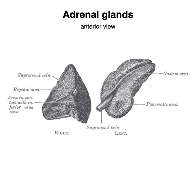
The adrenal glands are located superior and anteromedial to the kidneys, within the perirenal space, and enclosed by perirenal fascia. Each has a body and two limbs: a medial and lateral limb. Coronally, the right adrenal gland is more pyramidal shaped, while the left more is more crescentic 7. Axially, the right adrenal gland is linear or V-shaped with larger medial limb and smaller lateral limb, while left adrenal gland is triangular shaped1,11.
The right adrenal gland has a maximum width of 6.1 mm and the left adrenal gland has a maximum width of 7.9 mm 6. The cranio-caudal extent of the adrenal gland is less than 4 cm 11. Proportionately, the adrenal size is larger in neonates and infants, being almost one-third of the size of the kidney 2-4.
The adrenal gland consists of two portions: an outer cortex and an inner medulla. The outer cortex is derived by mesoderm while the inner medulla is derived from neural crest cells 11. The gland is covered by a fibrous capsule.
The cortex is subdivided into three zones (from outer to inner) 5,7:
zona glomerulosa
zona fasciculata
zona reticularis
The medulla (chromaffin cells) produces catecholamines (adrenaline, noradrenaline) and dopamine.
Locations
The adrenal glands lie superior and anteromedial to the kidneys 1. The left adrenal gland lies more anteromedial than superior when compared to the right adrenal gland 1. The right adrenal gland lies in the angle between the right lobe of liver and crus of the right diaphragm, behind the inferior vena cava 1. Meanwhile, the left adrenal gland lies posterolateral to the abdominal aorta 1. The right adrenal gland is lower and more medial to the spine than the left adrenal gland 11.
Relations
Right adrenal gland
medial: right crus of the diaphragm, right inferior phrenic artery
lateral: right lobe (bare area) of the liver
anterior: inferior vena cava (IVC)
posterior: right kidney 3,6,7
Left adrenal gland
medial: left crus of the diaphragm, left inferior phrenic artery
lateral: spleen
anterior: splenic artery, pancreas, stomach, lesser sac
posterior: left kidney 3,6,7
Arterial supply
The arterial supply is via three adrenal arteries 5:
-
superior adrenal artery which arises from the inferior phrenic artery
usually a group of 6-8 arteries arise 10
-
middle adrenal artery which arises from the abdominal aorta
often the largest of the adrenal arteries
inferior adrenal artery which arises from the renal artery
Venous drainage
-
venous drainage 5
-
adrenal veins emerge from the hilum and drain to different veins depending on the side:
-
left adrenal vein drains to the left renal vein
may form a common trunk with the inferior phrenic vein before entering the left renal vein
right adrenal vein drains to the IVC
-
-
Lymphatic drainage
lymphatics are contained within the capsule and drain to the para-aortic nodes 7,9
Innervation
presynaptic sympathetic fibres from thoracic splanchnic nerves synapse directly with chromaffin cells 9
postsynaptic fibres from coeliac, aorticorenal, and renal ganglia innervate surrounding vessels
Variant anatomy
-
takes on a flattened appearance
often in the presence of a pelvic kidney or renal agenesis
adrenal gland hypoplasia or agenesis 8
-
accessory adrenal rests
often near adrenal glands but may be found anywhere in the abdomen, pelvis or scrotum 9
Radiographic features
Generally, the size of the adrenal gland should not be thicker than the adjacent crus of diaphragm 11.
Ultrasound
-
anterior transverse scanning is the best approach but the adrenal glands are often difficult to see 6:
left adrenal gland seen ~40% at least once during scan
right adrenal gland seen ~80% at least once during scan
-
left adrenal gland is more difficult to visualise than the right because it is often posterior to the stomach and obscured by gas; this can be overcome by 6
intercostal scanning in the posterior axillary line
scanning through the spleen and left kidney with the left side of the patient elevated
CT
Normal adrenal glands have similar density with liver and spleen on plain CT scan 12.
The adrenal glands enhance after contrast administration to approximately 50-60 HU. Hyperenhancement of the adrenal gland is concerning for hypovolaemic shock.
MRI
Adrenal glands are more easily seen on MRI than on CT because surrounding fat that give them contrast. Adrenal glands are isointense or hypointense when compared to liver on both T1-weighted and T2-weighted images. On fat suppression, the adrenal glands lose signals depending on cholesterol content on the adrenal cortex 11.
Development
Adrenal glands develop in the peritoneum and descend, in contrast to kidneys that develop in the pelvis and ascend. If the kidneys failed to develop, the adrenal glands are still found in their expected positions but their shape may be more discoid due to lack of moulding by the kidneys 11.
At birth, the adrenal glands sizes are 1/3 of the kidneys. This gets smaller into adulthood where it becomes 1/13 of the kidneys in adults 11.
Related pathology
-
gamuts
-
vascular
-
traumatic
-
neoplastic
-
infections
-
inflammation
-
others


 Unable to process the form. Check for errors and try again.