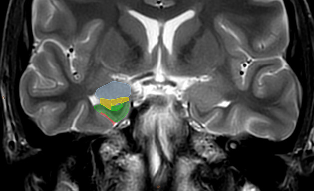
Amygdala
The amygdala is a very well studied part of the limbic system and forms part of the mesial temporal lobe.
Gross anatomy
The amygdala is a complex gray matter structure located anterior and superior to the temporal horn of the lateral ventricle and head of the hippocampus. The anterior extension of the amygdala is identified by the entorhinal sulcus.
Both the amygdala and the hippocampus are derived from the telencephalon. In 1997, Gloor classified the amygdala into three large groups of nuclei 2,3:
- basolateral
- corticomedial
- central group
Connections
Output fibers
The output fibers of the amygdala is mainly through two efferent fiber systems:
- stria terminalis
- ventral amygdalofugal pathway
The stria terminalis has a semicircular course in the body of the lateral ventricle. The level of the anterior commissure is a very important anatomical landmark because the stria terminalis splits into three parts:
- pre-commissural part
- commissural part
- post-commissural part
The pre-commissural fibers end in the septal region, and the post-commissural fibers end in the nucleus of stria terminalis. The commissural fibers connect the contralateral amygdala via the anterior commissure.
The ventral amygdalofugal pathway arises from the dorsomedial aspect of the amygdala, and courses medially through substantia innominata, anterior perforated substance, and fibers spread into the cerebral cortex, brainstem, mediodorsal thalamus, and hypothalamus. Direct efferent fibers project to the entorhinal cortex as well as the hippocampus 2.
Input fibers
The input fibers to the amygdala are mainly represented by sensory fibers, which transfer information from numerous subcortical regions such as the hypothalamus, basal ganglia, septal region, and autonomic centers in the brainstem. Both the ventral amygdalofugal pathway and the stria terminals carry afferent and efferent fibers together. Some input fibers project to the amygdala along the olfactory stria, and directly from the temporal lobe.
Reciprocal fibers
The basal nucleus of Meynert, and the nucleus of the diagonal band project reciprocally to the amygdala along the ventral amygdalofugal pathway 1,2.
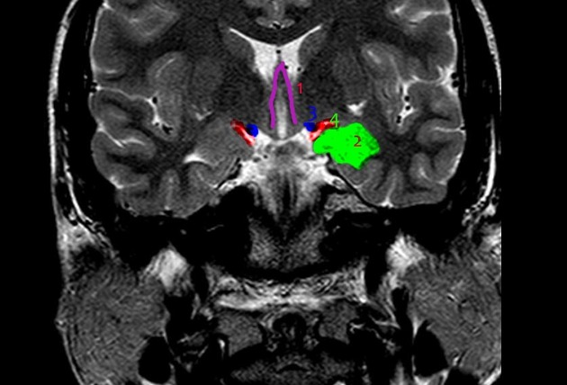
Relations
- superiorly: partly continuous with the inferomedial aspect of the claustrum
- laterally: optic tract
- posteriorly: hippocampus, connected to the tail of the caudate nucleus
Etymology
Amygdala is derived from the Greek amugdalē, meaning almond, as the complex is almond-shaped.
Related Radiopaedia articles
Anatomy: Brain
-
brain
- grey matter
- white matter
-
cerebrum
-
cerebral hemisphere (telencephalon)
- cerebral lobes and gyri
- frontal lobe
- parietal lobe
-
occipital lobe
- occipital pole
- lingual gyrus
- fusiform gyrus (Brodmann area 37)
- calcarine (visual) cortex
- cuneus
-
temporal lobe
- temporal pole
- temporal gyri
- mesial temporal lobe
- lateral parietotemporal line
- basal forebrain
- limbic system
- insula
-
cerebral sulci and fissures (A-Z)
- calcarine fissure
- callosal sulcus
- central (Rolandic) sulcus
- cingulate sulcus
- collateral sulcus
- inferior frontal sulcus
- inferior occipital sulcus
- inferior temporal sulcus
- interhemispheric fissure
- intraparietal sulcus
- lateral (Sylvian) sulcus
- lateral occipital sulcus
- marginal sulcus
- occipitotemporal sulcus
- olfactory sulcus
- paracentral sulcus
- paraolfactory sulcus
- parieto-occipital fissure
- posterior parolfactory sulcus
- precentral sulcus
- preoccipital notch
- postcentral sulcus
- rhinal sulcus
- rostral sulcus
- subparietal sulcus
- superior frontal sulcus
- superior occipital sulcus
- superior temporal sulcus
- cortical histology
- cerebral lobes and gyri
- white matter tracts
- deep grey matter
-
pituitary gland
- posterior pituitary and stalk (part of diencephalon)
- anterior pituitary
- inferior hypophyseal arterial circle
- diencephalon
-
cerebral hemisphere (telencephalon)
-
brainstem
- midbrain (mesencephalon)
- pons (part of metencephalon)
- medulla oblongata (myelencephalon)
- white matter
- grey matter (non-cranial nerve)
-
cerebellum (part of metencephalon)
- vermis
- cerebellar hemisphere
- cerebellar peduncles
- cerebral meninges (meninx primitiva)
- CSF spaces
-
cranial nerves (mnemonic)
- olfactory nerve (CN I)
- optic nerve (CN II)
- oculomotor nerve (CN III)
- trochlear nerve (CN IV)
- trigeminal nerve (CN V) (mnemonic)
- abducens nerve (CN VI)
- facial nerve (CN VII) (segments mnemonic | branches mnemonic)
- vestibulocochlear nerve (CN VIII)
- glossopharyngeal nerve (CN IX)
- vagus nerve (CN X)
- spinal accessory nerve (CN XI)
- hypoglossal nerve (CN XII)
- functional neuroanatomy
- CNS development
- cerebral vascular supply
- arteries
- vascular territories
-
circle of Willis
- internal carotid artery (ICA) (segments)
- vertebral artery
-
normal variants
- intracranial arterial fenestration
- internal carotid artery (ICA)
- anterior cerebral artery (ACA)
- middle cerebral artery (MCA)
- posterior cerebral artery (PCA)
- basilar artery
- persistent carotid-vertebrobasilar artery anastomoses (mnemonic)
-
cerebral venous system
- dural venous sinuses
-
cerebral veins
-
superficial veins of the brain
- superior cerebral veins (superficial cerebral veins)
- inferior cerebral veins
- superficial middle cerebral vein
- superior anastomotic vein (of Trolard)
- inferior anastomotic vein (of Labbe)
- deep veins of the brain
-
superficial veins of the brain
- normal variants
- arteries
- glymphatic pathway