
Antenatal soft markers on ultrasound
Citation, DOI, disclosures and article data
At the time the article was created Yuranga Weerakkody had no recorded disclosures.
View Yuranga Weerakkody's current disclosuresAt the time the article was last revised Arlene Campos had no financial relationships to ineligible companies to disclose.
View Arlene Campos's current disclosures- Soft antenatal sonographic markers
- Soft markers on antenatal ultrasound
- Soft signs in antenatal ultrasound
Antenatal soft ultrasound markers are fetal sonographic findings that are generally not abnormalities as such but are indicative of an increased age-adjusted risk of an underlying fetal aneuploidic or certain non-chromosomal abnormalities.
Most of the described features do not constitute a structural defect and may be detected on antenatal screening, typically during a second trimester (morphology) scan. Many are transient.
What is included or not appears to depend on the respective authorities in each region, and remains dynamic, with certain markers periodically emerging, or conversely losing favour.
The following are markers in common use at the time of writing. The list includes features having an association with:
An increased risk of aneuploidy and in some cases non-chromosomal abnormalities
increased nuchal thickness: >6 mm
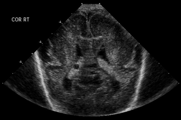
fetal ventriculomegaly: >10 mm
hypoplastic/absent nasal bone
-
shortened fetal long bones: less than 3rd centile for gestational age
An increased risk of non-chromosomal abnormalities when seen in isolation
fetal pyelectasis/fetal renal pelvic dilatation: antero-posterior diameter of the renal pelvis >4-5 mm
Soft markers of undefined association
Correct interpretation of these often requires correlation with other risk factors such as history, maternal age, and maternal serological results.
References
- 1. Van den hof MC, Wilson RD. Fetal soft markers in obstetric ultrasound. J Obstet Gynaecol Can. 2005;27 (6): 592-636. - Pubmed citation
- 2. Raniga S, Desai PD, Parikh H. Ultrasonographic soft markers of aneuploidy in second trimester: are we lost? MedGenMed. 2006;8 (1): 9. MedGenMed (link) - Free text at pubmed - Pubmed citation
- 3. Wax JR, Cartin A, Pinette MG et-al. Does the frequency of soft sonographic aneuploidy markers vary by fetal sex? J Ultrasound Med. 2005;24 (8): 1059-63. J Ultrasound Med (full text) - Pubmed citation
- 4. Agathokleous M, Chaveeva P, Poon LC et-al. Meta-analysis of second-trimester markers for trisomy 21. Ultrasound Obstet Gynecol. 2013;41 (3): 247-61. doi:10.1002/uog.12364 - Pubmed citation
Incoming Links
- Fetal clenched hands
- Rocker bottom foot
- Quadruple screening test
- Antenatal screening
- Shortened fetal long bones
- Echogenic fetal bowel
- Antenatal features of Down syndrome
- Echogenic intracardiac focus
- Hypoplastic nasal bone
- Shortened fetal femur
- Strawberry skull
- Fetal ventriculomegaly
- Clinodactyly
- Sandal gap deformity
- Choroid plexus cyst (antenatal)
Related articles: Pathology: Genitourinary
- obstetrics
-
first trimester
- ultrasound findings in early pregnancy
- embryo/fetus
- beta-hCG levels
- confirming intrauterine gestation
- pregnancy of unknown location (PUL)
- first trimester vaginal bleeding
- early structural scan
- aneuploidy testing
-
second trimester
- fetal biometry
- amniotic fluid volume
- fetal morphology assessment
- soft markers
- amnioreduction
- Doppler ultrasound
- nuchal translucency
- 11-13 weeks antenatal scan
- chorionic villus sampling (CVS) and amniocentesis
- other
- placenta
- placental anatomy
- placental developmental abnormalities
- placenta praevia
- spectrum of abnormal placental villous adherence
- abnormalities of cord insertion
- abruptio placentae
- placental pathology
- vascular pathologies of placenta
- placental infections
- placental masses
- molar pregnancy
- twin placenta
- miscellaneous
-
first trimester
- gynaecology
- acute pelvic pain
- chronic pelvic pain
- uterus
- ovaries
- ovarian follicle
- ovarian torsion
- pelvic inflammatory disease
- ovarian cysts and masses
- paraovarian cyst
- polycystic ovaries
- ovarian hyperstimulation syndrome
- post-hysterectomy ovary
- cervix
- fallopian tube
- other
- male genital tract
- prostate gland
- transrectal ultrasound
- prostate tumours
- infections of the prostate
-
prostatitis
- acute bacterial prostatitis
-
chronic prostatitis
- chronic bacterial prostatitis
- chronic prostatitis and chronic pelvic pain syndrome (CPPS)
- asymptomatic inflammatory prostatitis
- granulomatous prostatitis
- emphysematous prostatitis
- prostatic abscess
-
prostatitis
- benign prostatic hypertrophy
- cystic lesions of the prostate
- prostatic calcification
- prostatic infarction
- testes
-
unilateral testicular lesion
- testicular torsion
- orchitis
- testicular trauma
-
germ cell tumours of the testis
- testicular seminoma
-
non seminomatous germ cell tumours
- mixed germ cell tumour
- yolk sac tumour (endodermal sinus tumour)
- embryonal cell carcinoma
- choriocarcinoma
- testicular teratoma
- testicular epidermoid (teratoma with ectodermal elements only)
- burned out testis tumour
- sex cord / stromal tumours of the testis
- testicular cyst
- testicular lymphoma
- bilateral testicular lesion
- paratesticular lesions
- epididymis
- other
- polyorchidism
- cryptorchidism
- tubular ectasia of the rete testis
- cystadenoma of the rete testis
- testicular sarcoidosis
- testicular tuberculosis
- spermatic cord
- fibrous pseudotumour of the scrotum
- scrotal leiomyosarcoma
- testicular adrenal rest tumours (TARTs)
- tunica vaginalis testis mesothelioma
- splenogonadal fusion
- testicular vasculitis
- abnormal testicular Doppler flow (differential)
-
unilateral testicular lesion
- penis
- prostate gland
- KUB
- kidneys
- normal renal anatomy
- hydronephrosis
- urolithiasis
- renal masses
- renal cystic disease
- renal infection
- vascular
- trauma
- ureter
- normal ureter anatomy
- ureteral stricture
- ureteral dilatation
- ureteral anomalies
- ureteral tumours
- ureteral trauma
- other
- bladder
- kidneys


 Unable to process the form. Check for errors and try again.