
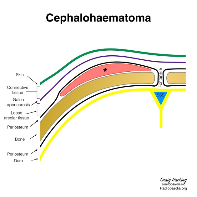
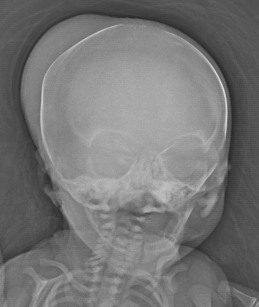
Cephalohematomas are traumatic subperiosteal hematomas of the skull that are usually caused by birth injury. They are bound between the periosteum and cranium, and therefore cannot cross sutures. Being bound by a suture line distinguishes them from subgaleal hematoma, which can cross sutures.
On this page:
Epidemiology
Cephalohematomas are as prevalent as 1-2% in spontaneous vaginal deliveries and 3-4% in forceps or vacuum-assisted deliveries. The incidence increases with ventouse and forceps extraction and thus more common in primiparous mothers. There may be a greater male predilection 4.
Clinical presentation
Cephalohematomas tend to increase in size following birth and will present as a firm mass. If blood loss is severe, infants may develop jaundice, anemia, and hypotension 6.
Radiographic features
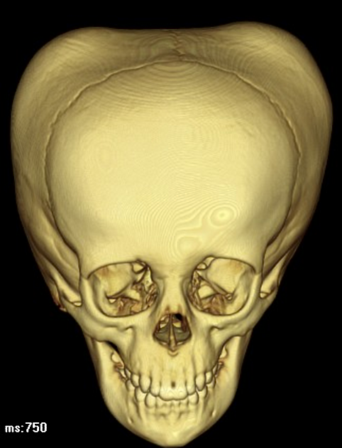
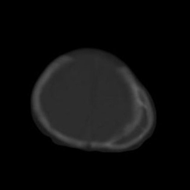
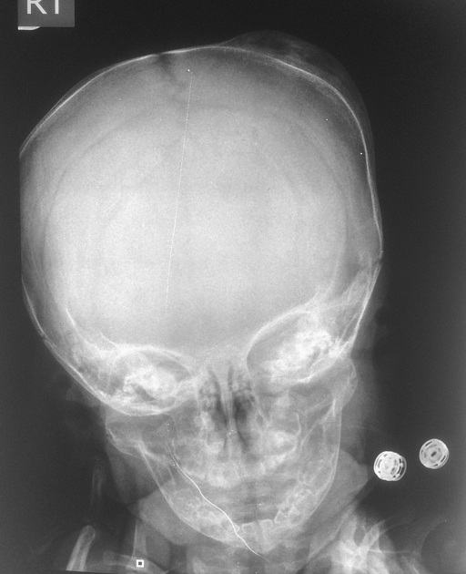
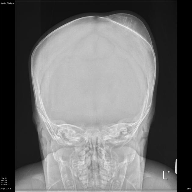
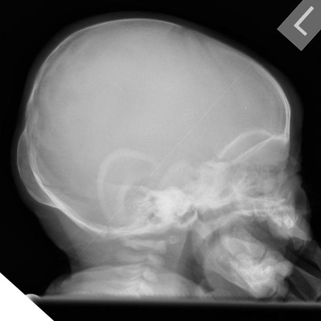
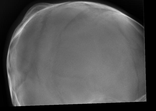
Cephalohematomas are clinically diagnosed and infrequently imaged. They can be unilateral or bilateral, and appear as subgaleal fluid collections bounded by suture lines. In the setting of craniosynostosis, the blood products are able to traverse the affected suture 5. By 2-3 weeks, they may become peripherally calcified 5. When calcified there is a hard swelling with a comparatively soft center that may clinically give the appearance of a depressed skull fracture 6.
Ultrasound
separation of the scalp by subperiosteal hemorrhage of moderate echogenicity 5
underlying brain is usually normal 5
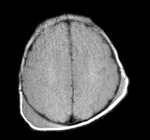
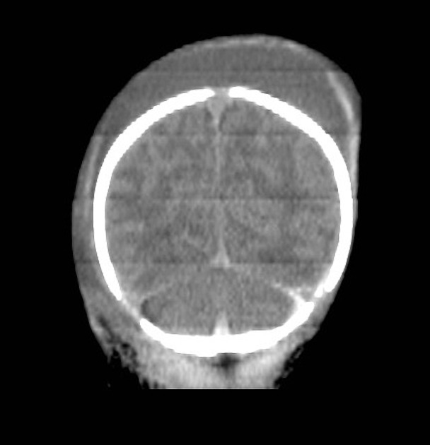
CT
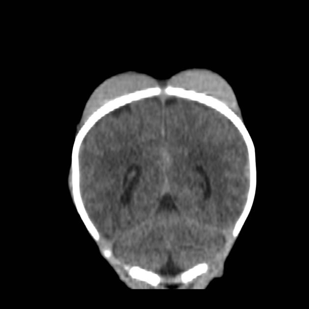
acute cephalohematoma will appear as a crescent shape adjacent to the external table of the skull 6
hyperdense if calcified
resolving lesions can appear concerning with regions of erosive change and periosteal reaction 6
MRI
acute cephalohematoma will appear as a crescent shape adjacent to the external table of the skull 6
hyperintensity on T1 and T2 weighted imaging in cases of subacute presentation but appearance will vary according to the age of the hematoma 6, and presence of calcium
Complications
Treatment and prognosis
Most resolve spontaneously in weeks but can take a maximum of 3-4 months 6.


 Unable to process the form. Check for errors and try again.