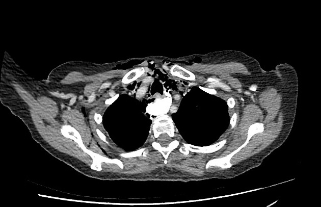
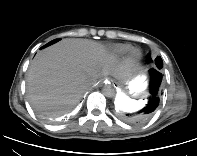
CT esophagography is a CT study designed to primarily evaluate the esophagus, particularly in the situation of esophageal trauma and potential perforation. It has been developed partly as an alternative to fluoroscopic barium swallow evaluation in this situation.
On this page:
Indications
potential esophageal perforation
evaluation of potential esophageal anastomotic leak
Technique
The are multiple potential ways to perform a CT esophagram, with the key variables:
-
type of oral contrast to give
it is not clear whether diatrizoate meglumine (Gastrografin) or a solution of iodinated low-osmolar contrast medium (e.g. Omnipaque) is better; if the patient is an aspiration risk, then avoiding a high-osmolar contrast agent such as Gastrografin seems prudent
protocols do not include the use of barium sulfate contrast medium
-
how much oral contrast to give
both diluted and undiluted contrast formulations have been used in protocols without a clear consensus
how quickly to begin scanning
whether to administer IV contrast
One such protocol 1:
scan field of view from the angle of the mandible to the level of the iliac crests
a non contrast series is obtained
the patient then drinks half a cup (~4 oz) of water soluble contrast agent
this is then followed with the patient drinking the other half of the cup in a supine position (with a straw)
after a 5 second delay, the post swallow series is obtained
could be repeated in the prone position if an anterior leak is suspected on the non contrast series
Contraindications
allergy to the oral contrast agent
-
an obtunded/comatose patient, since the patient cannot protect his or her airway
in select situations, a nasogastric tube could be advanced and enteric contrast administered through it before scanning


 Unable to process the form. Check for errors and try again.