
Cytomegalovirus (CMV) pneumonia is a type of viral pneumonitis and occurs due to infection with cytomegalovirus (CMV), which is a member of the Herpetoviridae family.
On this page:
Epidemiology
Cytomegalovirus infection is particularly important in those who are immunocompromised (e.g. those with AIDS, allogeneic bone marrow transplantation recipient 3). In recipients of hematopoietic stem cell transplantations, the incidence of CMV can be around 20-35% 8.
Pathology
A major biological characteristic of cytomegalovirus (as with other herpes viruses) is its ability to become latent in the human host and therefore the potential for reactivation.
Radiographic features
Plain radiograph
Findings on chest radiographs are usually non-specific.
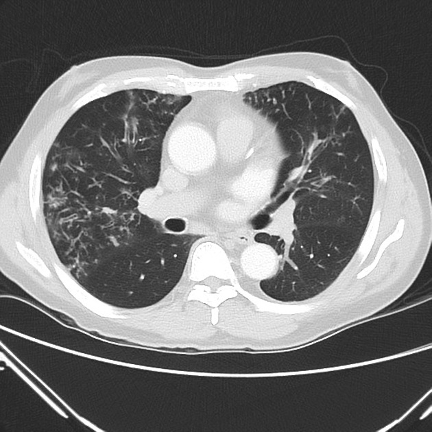
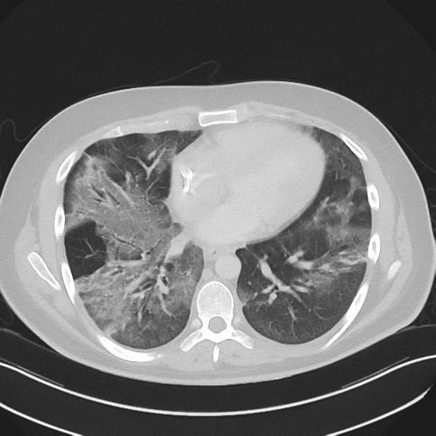
CT
CT findings are non-specific and diverse and have been described without distinction between AIDS and non-AIDS patients. Commonly described findings include:
-
mixed alveolar-interstitial infiltrative opacification
-
relatively common feature, may be seen in ~67% of cases 5
-
-
small pulmonary nodules 4,7
nodules tend to have bilateral symmetrical distribution and involve all zones 6
-
confluent consolidation 1
may be more marked towards the lower lobes 6
interstitial reticulation without air space opacification 1,4
Other described features include:
pulmonary masses 4
tree-in-bud changes 5
Differential diagnosis
The imaging differential is broad but in the immunosuppressed population consider:
-
pneumocystis jirovecii pneumonia (PCP) 8
may contain (not always) small intrapulmonary cysts on CT
may have a more apical distribution 8
the ground glass changes may be more homogeneous 8
other forms of viral pneumonitis


 Unable to process the form. Check for errors and try again.