
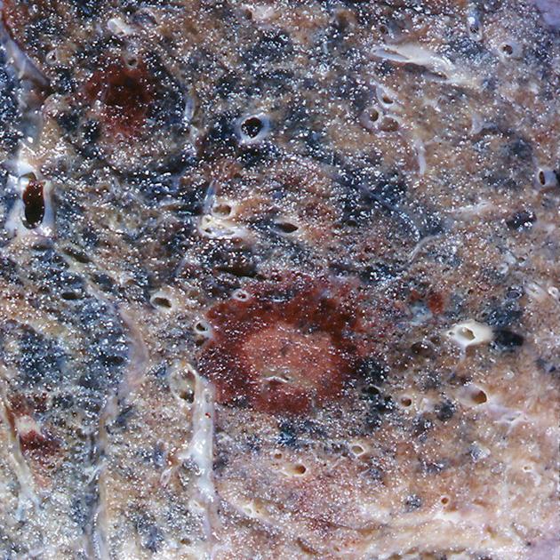
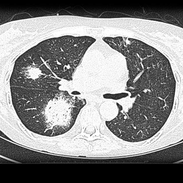
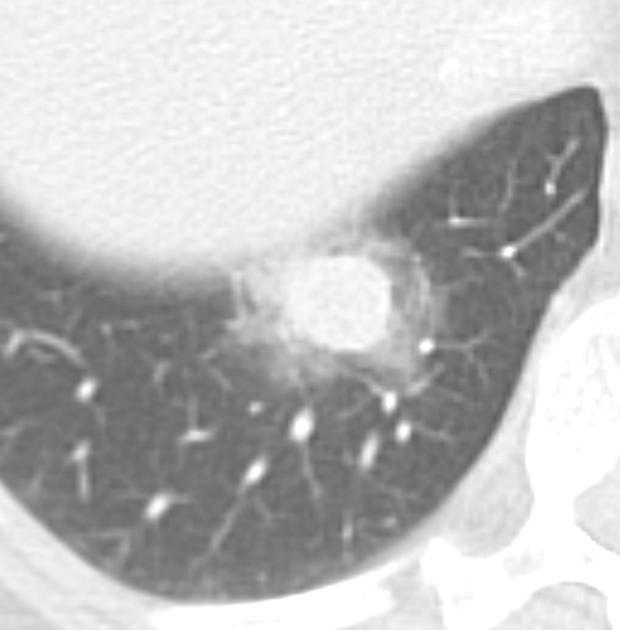
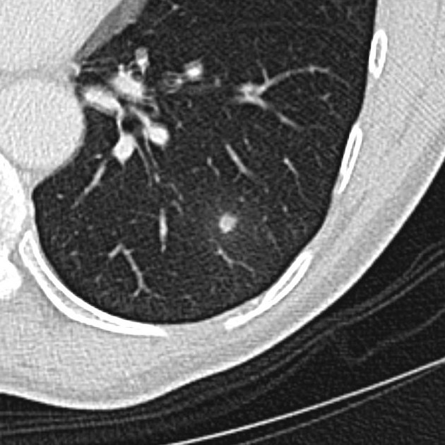
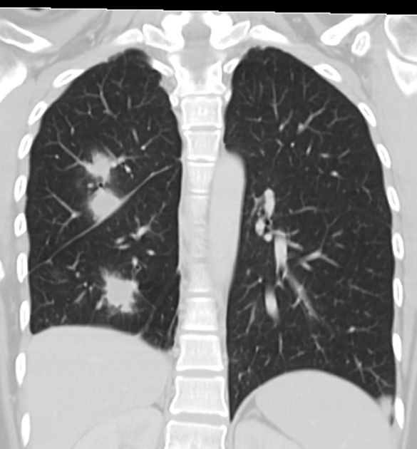
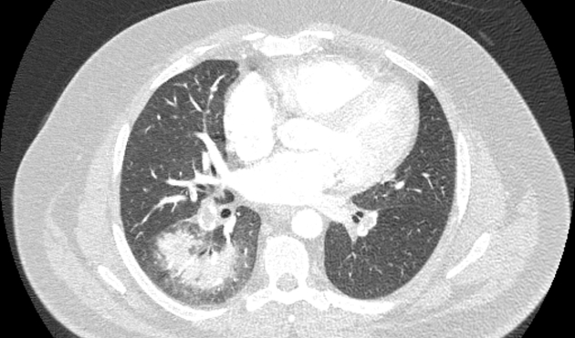
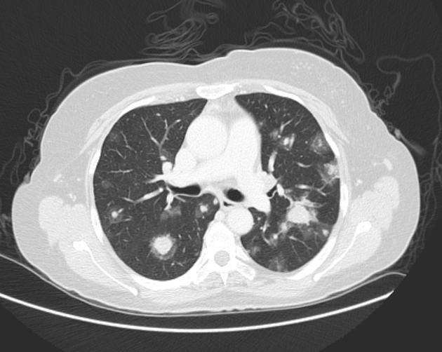
The halo sign in chest imaging is a feature seen on lung window settings, ground glass opacity surrounding a pulmonary nodule or mass and represents hemorrhage. It is typically seen in angioinvasive aspergillosis.
On this page:
Pathology
Histopathologically, it represents a focus of pulmonary infarction surrounded by alveolar hemorrhage.
Diagnostic considerations
Other entities that may give a halo sign include:
Infectious disease
- fungi
- septic embolism
- mycobacterial
- rickettsia - rickettsia pneumonia
- Coxiella burnetii - Q fever pneumonia
- viral: herpes simplex virus, varicella zoster virus (chickenpox), cytomegalovirus, myxovirus
Malignancy
- primary tumors
- adenocarcinoma of the lung, adenocarcinoma in situ or minimally invasive (formerly bronchioalveolar carcinoma) - has been described as the most commonly encountered with halo sign in immunocompetent patients 5
- squamous cell carcinoma of the lung
- Kaposi sarcoma
- pulmonary lymphoma
-
lung metastases (especially - hemorrhagic pulmonary metastases):
- angiosarcoma
- choriocarcinoma
- osteosarcoma
- melanoma
- metastasis from gastrointestinal adenocarcinoma (<10%)
Non-neoplastic, non-infectious, inflammatory diseases
- pulmonary infarction
- granulomatosis with polyangiitis
- eosinophilic lung disease
- pulmonary endometriosis
- organizing pneumonia
- hypersensitivity pneumonitis
- iatrogenic injury
- pulmonary pseudoaneurysm
See also
- halo sign (breast)
- halo sign (ultrasound)
- reversed halo sign (atoll sign)


 Unable to process the form. Check for errors and try again.