
This is a basic article for medical students and other non-radiologists
Intracranial tumors comprise a heterogeneous group of tumors. In adult patients, the majority represent metastatic disease with a smaller proportion being primary brain tumors. Metastasis to the brain occurs, most commonly, from lung, breast, melanoma, renal cell, and colorectal cancers.
On this page:
Reference article
This is a summary article; read more in our article on intracranial tumors.
Summary
-
epidemiology
incidence increases with age
equivocal gender distribution
-
risk factors
malignancy elsewhere
-
presentation
headache
-
features of raised intracranial pressure
nausea & vomiting worse in the morning or positional
altered mental state
focal neurology may occur as the tumor grows
adult-onset seizures
-
incidental finding
some tumors may not cause symptoms
patients may be imaged for another reason, e.g. trauma
-
pathophysiology
-
heterogeneous group of tumors
metastases, e.g. lung, breast, renal
meningiomas
primary parenchymal tumors
pituitary or pineal tumors
cranial nerve schwannomas
tumors are graded using the WHO grade
-
-
investigation
CT is often the first test performed to assess presenting symptoms
MRI may be used with symptoms of headaches or seizures
MRI is the investigation of choice to characterize the tumor
-
treatment
parenchymal brain tumors generally have a poor prognosis
-
treatment should be in specialist centers
steroids may alleviate symptoms caused by edema
antiseizure medications may help for those with seizures
a biopsy may be performed neurosurgically
some tumors may be removed, e.g. pituitary tumors
stereotactic radiotherapy can be used for small lesions
Imaging
-
role of imaging
confirm intracranial abnormality and prioritise MRI
tumor characterization
help to determine the grade, and make a decision about biopsy
follow up
-
radiographic features
-
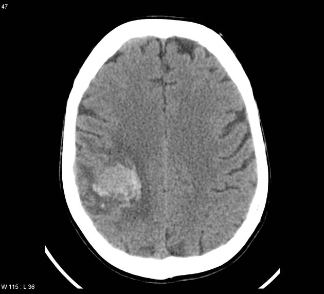
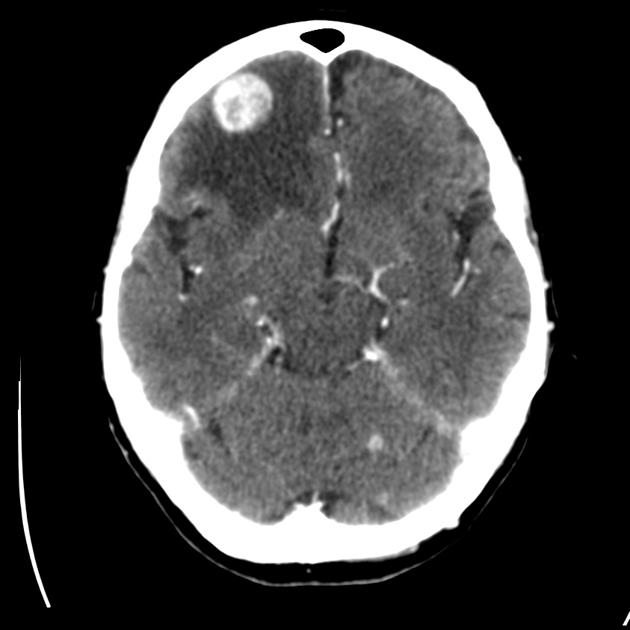
CT
often the first line test
variety of appearances depending on the tumor
hypo- or hyperdense, irregular, well-defined, peripheral or deep
useful to determine edema and mass effect
contrast may make lesions more conspicuous
CT is especially helpful for determining bony involvement
-
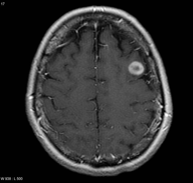
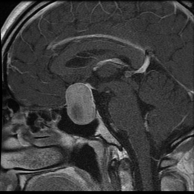
MRI
investigation of choice
fantastic contrast and spatial resolution
origin of tumors can be determined
different sequences are used to determine the likely diagnosis
specialized sequences can be useful to look at tumor metabolites
-


 Unable to process the form. Check for errors and try again.