
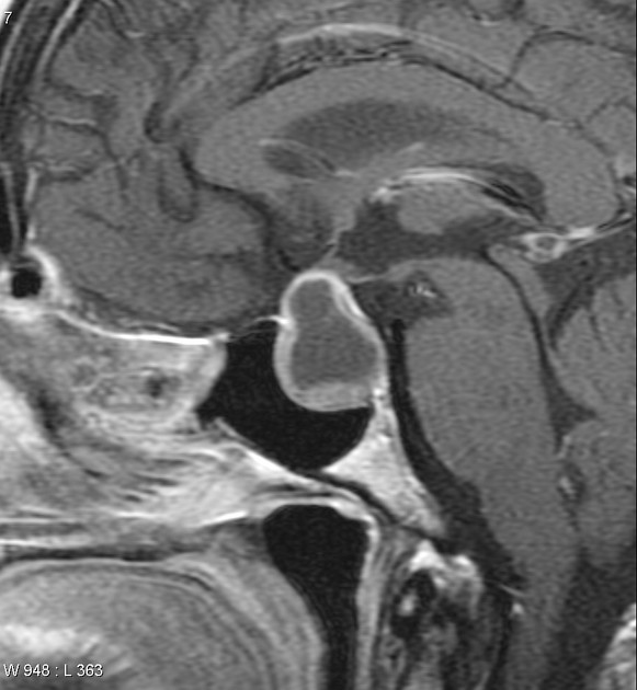
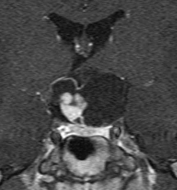
Mixed cystic and solid pituitary region mass
Citation, DOI, disclosures and article data
Citation:
Bell D, Siriwardana D, Sharma R, et al. Mixed cystic and solid pituitary region mass. Reference article, Radiopaedia.org (Accessed on 18 Feb 2025) https://doi.org/10.53347/rID-17449
Permalink:
rID:
17449
Article created:
Disclosures:
At the time the article was created Frank Gaillard had no recorded disclosures.
View Frank Gaillard's current disclosures
Last revised:
Disclosures:
At the time the article was last revised Daniel J Bell had no recorded disclosures.
View Daniel J Bell's current disclosures
Revisions:
3 times, by
3 contributors -
see full revision history and disclosures
Systems:
Sections:
Synonyms:
- Mixed solid and cystic pituitary region mass
- Mixed cystic and solid pituitary region masses
- Mixed solid and cystic pituitary region masses
A mixed cystic and solid pituitary region mass has a limited differential.
Differential diagnosis
-
craniopharyngioma
- both papillary (more solid) and adamantinomatous (more cystic)
-
macroadenoma
- cystic change / necrosis / previous hemorrhage
Most other solid and enhancing pituitary region masses can have cystic areas occasionally, but it is not usually a dominant/significant component.
References
- 1. Park, M., Lee, S., Choi, J., Kim, S., Kim, S., Shin, N., Kim, J. and Ahn, S. (2015). Differentiation between Cystic Pituitary Adenomas and Rathke Cleft Cysts: A Diagnostic Model Using MRI. American Journal of Neuroradiology, 36(10), pp.1866-1873.
Incoming Links
Related articles: Pituitary region masses
-
general reading
- pituitary gland anatomy
- pituitary MRI - an approach
-
pituitary region masses
- most common pituitary region masses
- solid and enhancing pituitary region mass
- mixed cystic and solid pituitary region mass
- mostly/purely cystic pituitary region masses
- purely intrasellar pituitary mass
- pituitary region mass with intrinsic high T1 signal
- abnormal enhancement/bulkiness of the pituitary infundibulum
- enlarged sella turcica
- mnemonic: SATCHMO
- history of imaging the pituitary region
-
pathology
-
pituitary tumors
- pituitary adenoma (commonest in the adult population)
- pituitary carcinoma
- pituitary lymphoma
- meningioma
- craniopharyngioma
- optic pathway glioma
- germinoma
- chordoma
- dermoid (CNS) / epidermoid / intracranial teratoma
- pituicytoma
- spindle cell oncocytomas
- pituitary metastases
- granular cell tumor of the pituitary (pituitary choristoma)
- pilocytic astrocytoma of the neurohypophysis (infundibuloma)
- cellular infiltrates
- other lesions
- anterior circulation berry aneurysm
- hamartoma (tuber cinereum hamartoma)
- Rathke cleft cyst
- intracranial lipoma
- sphenoid sinus mucocoele
- pituitary abscess
- pituitary stone
-
pituitary tumors


 Unable to process the form. Check for errors and try again.