
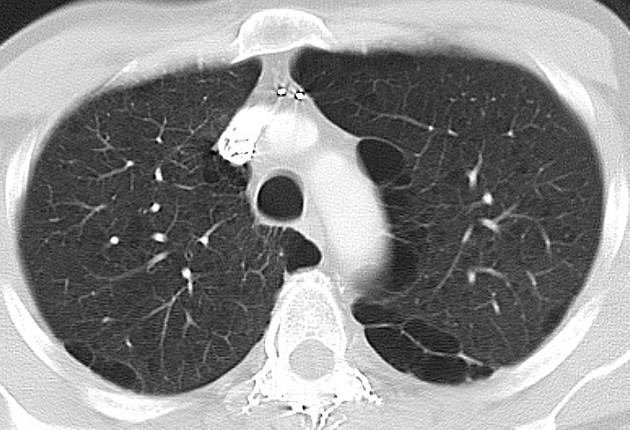
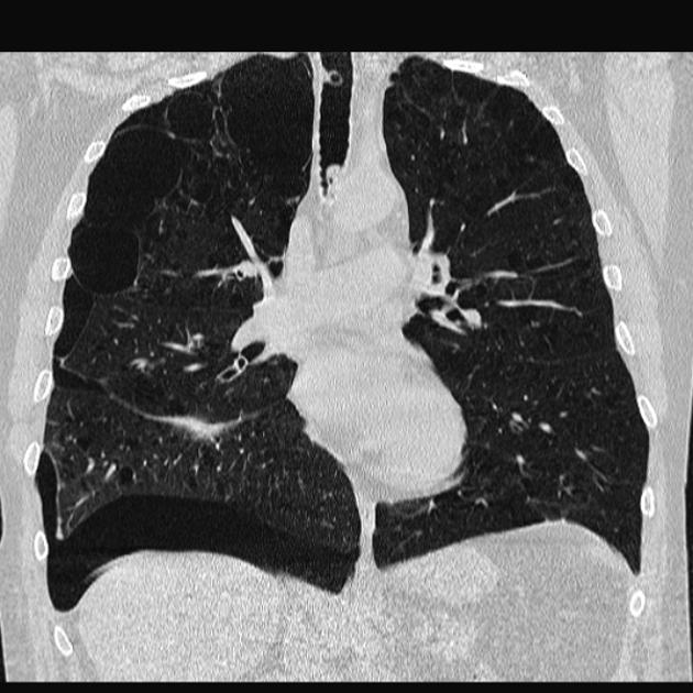
Pulmonary bullae (singular bulla) are gas-containing cystic structures formed by confluent destroyed and dilated airspaces (distal to terminal bronchioles).
On this page:
Terminology
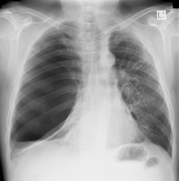
A giant bulla is arbitrarily defined as one that occupies at least one third of the volume of a hemithorax 5. See also Idiopathic giant bullous emphysema.
Pathology
Although bullae are typically larger than blebs, there is no defining size limit for either entity in the 2024 Fleischner glossary 6, although a size of >1 cm was defined in the 2008 Fleischner glossary 7. Large bullae can compress adjacent lung causing atelectasis.
They are often subpleural in location (see: subpleural bullae) and are typically larger in the apices which experience the greatest tension.
Etiology
The most common cause is smoking-related paraseptal emphysema or centrilobular emphysema.
Complications
infection
hemorrhage
Radiographic features
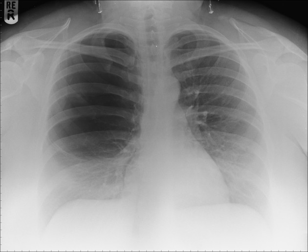
Bullae are typically well-demarcated and rounded with a fine smooth wall (almost imperceptible) composed of a thin layer of collapsed lung 6,8.
Differential diagnosis
when large, a bulla can mimic a pneumothorax


 Unable to process the form. Check for errors and try again.