
Scrotal haematocele
Citation, DOI, disclosures and article data
At the time the article was created Ian Bickle had no recorded disclosures.
View Ian Bickle's current disclosuresAt the time the article was last revised Mostafa Elfeky had no recorded disclosures.
View Mostafa Elfeky's current disclosures- Haematocoele
- Scrotal haematocoeles
- Scrotal haematocoele
- Scrotal haematoceles
- Scrotal hematocele
- Scrotal hematoceles
- Hematocele
- Haematocele
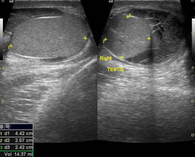
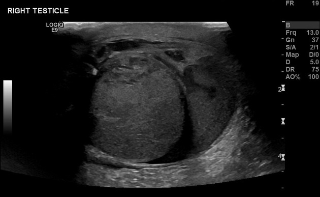
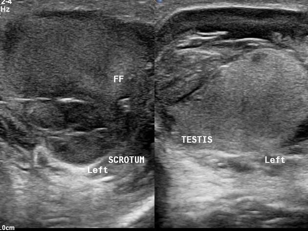
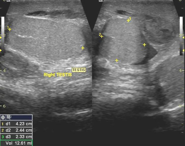
Scrotal haematoceles are collections of blood within the scrotal sac, but outside of the testis.
On this page:
Pathology
A haematocele normally results from trauma to the scrotum, or on occasion following surgery. Some think that a varicocele is a risk factor for developing a haematocele 4.
Radiographic features
Ultrasound
Ultrasound is usually the sole imaging modality used, typically being first line for those with a scrotal swelling or pain following trauma to the scrotum. Unlike a hydrocele, which is anechoic, a haematocele is of increased echogenicity and often has septa within. If the ultrasonographic investigation is performed right after the event, a fresh haematocele can appear anechoic, though some turbulence will occasionally be seen within it.
Differential diagnosis
A clinical differential would be for a scrotal wall haematoma.
See also
Quiz questions

References
- 1. Proctor RD, Tung K. Persistent scrotal lesion. Br J Radiol. 2009;82 (983): 966-7. Br J Radiol (full text) - doi:10.1259/bjr/31160455 - Pubmed citation
- 2. Saez F, Descalzo MJ, Herrera B et-al. Hematocele secondary to rupture of an abdominoscrotal hydrocele. Arch. Esp. Urol.66 (9): 877-9. Pubmed citation
- 3. Chaudhary S, Bhullar JS, Subhas G et-al. Hematocele after laparoscopic appendectomy. JSLS. 2012;16 (4): 660-2. doi:10.4293/108680812X13517013316717 - Free text at pubmed - Pubmed citation
- 4. Sommers D, Winter T. Ultrasonography evaluation of scrotal masses. Radiol. Clin. North Am. 2014;52 (6): 1265-81. doi:10.1016/j.rcl.2014.07.014 - Pubmed citation
Incoming Links
Related articles: Pathology: Genitourinary
- obstetrics
-
first trimester
- ultrasound findings in early pregnancy
- embryo/fetus
- beta-hCG levels
- confirming intrauterine gestation
- pregnancy of unknown location (PUL)
- first trimester vaginal bleeding
- early structural scan
- aneuploidy testing
-
second trimester
- fetal biometry
- amniotic fluid volume
- fetal morphology assessment
- soft markers
- amnioreduction
- Doppler ultrasound
- nuchal translucency
- 11-13 weeks antenatal scan
- chorionic villus sampling (CVS) and amniocentesis
- other
- placenta
- placental anatomy
- placental developmental abnormalities
- placenta praevia
- spectrum of abnormal placental villous adherence
- abnormalities of cord insertion
- abruptio placentae
- placental pathology
- vascular pathologies of placenta
- placental infections
- placental masses
- molar pregnancy
- twin placenta
- miscellaneous
-
first trimester
- gynaecology
- acute pelvic pain
- chronic pelvic pain
- uterus
- ovaries
- ovarian follicle
- ovarian torsion
- pelvic inflammatory disease
- ovarian cysts and masses
- paraovarian cyst
- polycystic ovaries
- ovarian hyperstimulation syndrome
- post-hysterectomy ovary
- cervix
- fallopian tube
- other
- male genital tract
- prostate gland
- transrectal ultrasound
- prostate tumours
- infections of the prostate
-
prostatitis
- acute bacterial prostatitis
-
chronic prostatitis
- chronic bacterial prostatitis
- chronic prostatitis and chronic pelvic pain syndrome (CPPS)
- asymptomatic inflammatory prostatitis
- granulomatous prostatitis
- emphysematous prostatitis
- prostatic abscess
-
prostatitis
- benign prostatic hypertrophy
- cystic lesions of the prostate
- prostatic calcification
- prostatic infarction
- testes
-
unilateral testicular lesion
- testicular torsion
- orchitis
- testicular trauma
-
germ cell tumours of the testis
- testicular seminoma
-
non seminomatous germ cell tumours
- mixed germ cell tumour
- yolk sac tumour (endodermal sinus tumour)
- embryonal cell carcinoma
- choriocarcinoma
- testicular teratoma
- testicular epidermoid (teratoma with ectodermal elements only)
- burned out testis tumour
- sex cord / stromal tumours of the testis
- testicular cyst
- testicular lymphoma
- bilateral testicular lesion
- paratesticular lesions
- epididymis
- other
- polyorchidism
- cryptorchidism
- tubular ectasia of the rete testis
- cystadenoma of the rete testis
- testicular sarcoidosis
- testicular tuberculosis
- spermatic cord
- fibrous pseudotumour of the scrotum
- scrotal leiomyosarcoma
- testicular adrenal rest tumours (TARTs)
- tunica vaginalis testis mesothelioma
- splenogonadal fusion
- testicular vasculitis
- abnormal testicular Doppler flow (differential)
-
unilateral testicular lesion
- penis
- prostate gland
- KUB
- kidneys
- normal renal anatomy
- hydronephrosis
- urolithiasis
- renal masses
- renal cystic disease
- renal infection
- vascular
- trauma
- ureter
- normal ureter anatomy
- ureteral stricture
- ureteral dilatation
- ureteral anomalies
- ureteral tumours
- ureteral trauma
- other
- bladder
- kidneys


 Unable to process the form. Check for errors and try again.