
A subchondral fracture is a fracture of the trabecular cancellous bone just beneath the subchondral bone plate without disruption of the articular surface 1.
On this page:
Epidemiology
Subchondral insufficiency fractures are more common in elderly women 1,4,6. Subchondral fractures due to trauma can occur at any age.
Risk factors
Associations
Associated injuries include 4-7:
ligamentous injuries
Clinical presentation
Patients will usually present with pain on weight-bearing in the affected joint, improving with rest 2.
Pathology
Subchondral fractures are usually a consequence of compressive forces, transmitted from the cartilage to the subchondral bone plate and from there to the trabeculae, which fail to resist that force and break or fracture 3. They can also occur in conjunction with twisting and ligamentous injuries.
Etiology
acute trauma 1
repetitive microtrauma/overuse fracture
Location
The weight-bearing joints such as the knee, hip, and ankle joints are more commonly affected.
Radiographic features
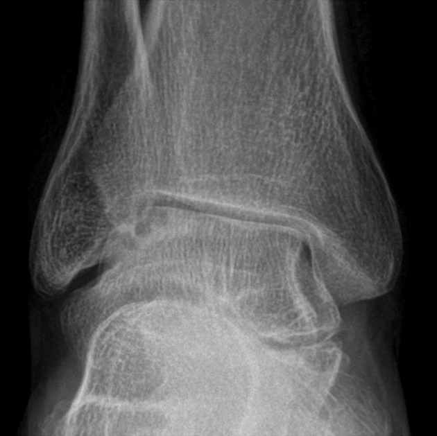
Usually, subchondral fractures present as linear or curvilinear structures often paralleling the subchondral bone plate, with or without areas of subchondral collapse 2.
Plain radiograph
Sometimes visible as subchondral hyperlucency with a decrease in bone density. Sclerotic lines as a result of impaction or as slight deformities of the joint line in case of subchondral collapse can sometimes be seen particularly at a later stage 4.
CT
directly visible subchondral trabecular discontinuity on high resolution
signs of subcortical impaction visible as subchondral hyperdensity
subchondral bone marrow edema on dual-energy CT
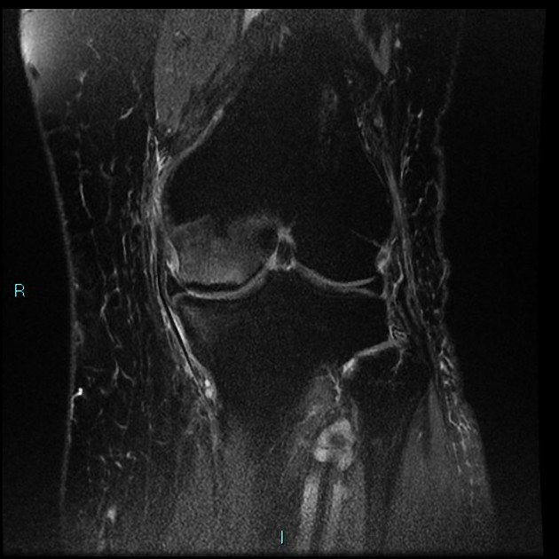
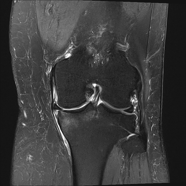
MRI
The fracture can be seen as irregular linear or curvilinear subchondral low signal intensity structure near the subchondral bone plate of low signal intensity in T1-weighted images and also sometimes, but not always in T2-weighted images 1,2,4-8. The area between the fracture line and the articular surface should be of high signal in T2 weighted images 2,4.
Almost always there will be associated bone marrow edema best appreciated in fat-saturated T2 weighted and intermediate or proton-density weighted images 4.
Signal characteristics
T1: low signal intensity fracture line, intermediate to a weak low signal of the surrounding stress reaction 2
T2: low signal intensity fracture line, but not always visible 5
IM/PDFS: high signal intensity stress reaction / bone marrow edema 2,5
Treatment and prognosis
Low-grade subchondral fractures in particular, if there is no collapse of the subchondral bone plate, can be treated conservatively with restricted weight-bearing 2,3 and non-steroidal anti-inflammatory drugs. Prostaglandin I-1 and/or bisphosphonates might be considered 5.
Joint preserving surgical treatments of subchondral fractures include microfracture, drilling, subchondroplasty 10, or in the hip joint: transtochanteric osteotomy 11.
High-grade fluid-filled lesions with cortical collapse and severe osteoarthritis might require arthroplasty.
Complications
They can progress to subchondral collapse, osteochondral injury, and osteochondral defects 1.
Differential diagnosis
-
osteonecrosis 2,5
concave, smooth half-moon/crescentic appearing necrotic segment
double line sign
serpiginous line in the chronic phase
-
also involves the cartilage
-
migratory transient bone marrow edema 2
no subchondral fracture line
-
subchondral cysts
usually in conjunction with chondral damage
-
usually affects all parts of the joint
-
usually in adolescents


 Unable to process the form. Check for errors and try again.