
Synovial plicae (knee)
Citation, DOI, disclosures and article data
Citation:
Gaillard F, Vadera S, Weerakkody Y, et al. Synovial plicae (knee). Reference article, Radiopaedia.org (Accessed on 22 Feb 2025) https://doi.org/10.53347/rID-9174
Permalink:
rID:
9174
Article created:
Disclosures:
At the time the article was created Frank Gaillard had no recorded disclosures.
View Frank Gaillard's current disclosures
Last revised:
Disclosures:
At the time the article was last revised Sonam Vadera had no recorded disclosures.
View Sonam Vadera's current disclosures
Revisions:
19 times, by
12 contributors -
see full revision history and disclosures
Systems:
Sections:
Synonyms:
- Plicae of the knee
- Plica of the knee
- Synovial plica of the knee
Synovial plicae are folds of synovium, thought to represent embryologic remnants. They are common, present in ~90% of arthroscopies 3.
They have been implicated in anterior knee pain and possibly in chondromalacia patellae although their role remains controversial 1,3.
On this page:
Gross anatomy
Some plicae have been described with multiple names in the literature representing the same structure 1,2:
- medial patellar plica: most common symptomatic plica
- suprapatellar plica / superior patellar plica
- infrapatellar plica / ligamentum mucosum: most common knee plica
- lateral patellar plica: rare (<1%)
They range in shape from a ridge or shelf to more discrete cord-like structure 3.
Radiographic features
CT arthrography
May be seen as a shelf like band.
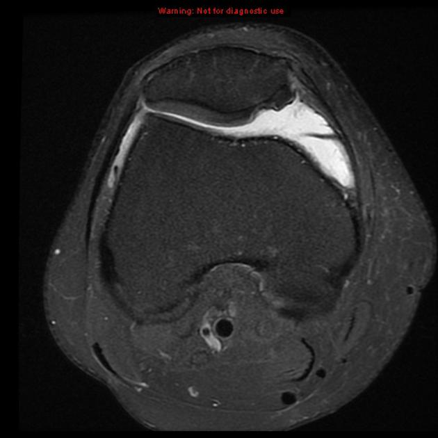
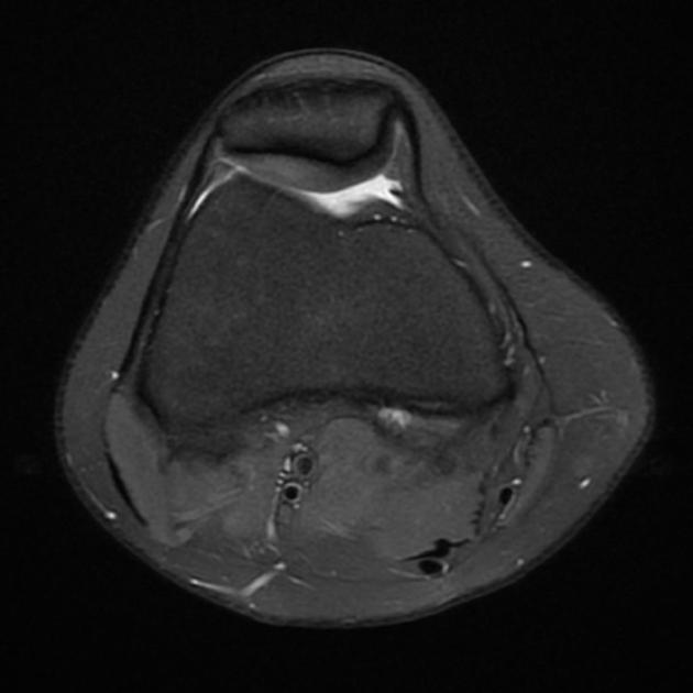
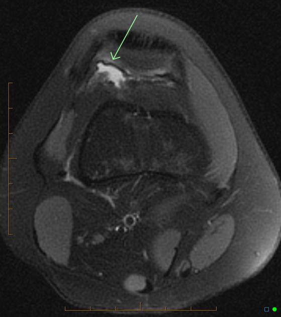
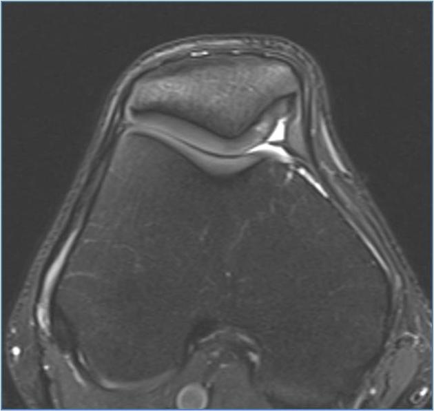
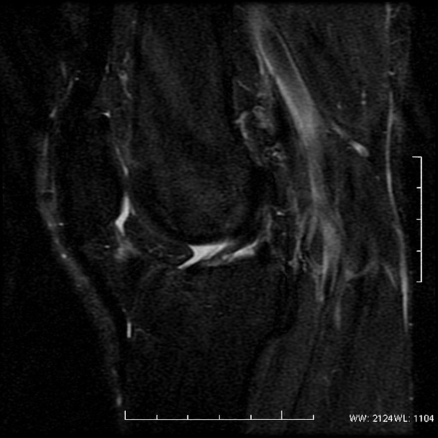
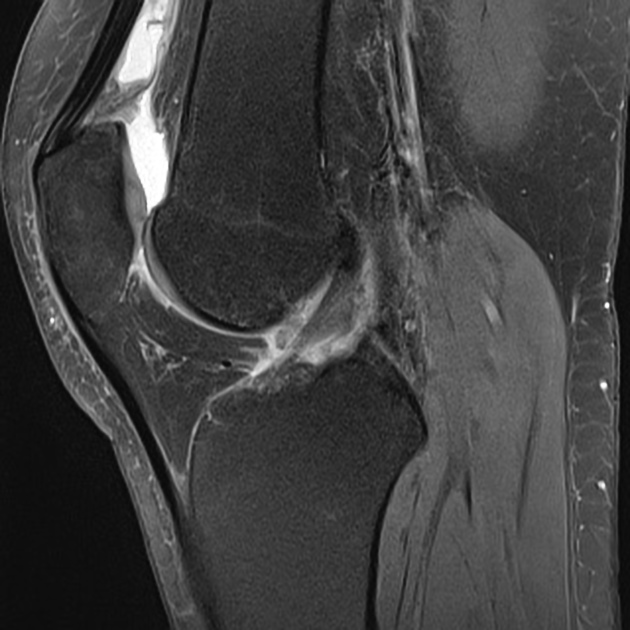
MRI
- PD/T2: typically seen as a band-like low signal structure traversing through the joint space (arthrography study or when joint effusion is present)
Related pathology
References
- 1. Hodge JC, Ghelman B, O'Brien SJ et-al. Synovial plicae and chondromalacia patellae: correlation of results of CT arthrography with results of arthroscopy. Radiology. 1993;186 (3): 827-31. Radiology (abstract) [pubmed citation]
- 2. García-valtuille R, Abascal F, Cerezal L et-al. Anatomy and MR imaging appearances of synovial plicae of the knee. Radiographics. 22 (4): 775-84. Radiographics (full text) - Pubmed citation
- 3. Boles CA, Martin DF. Synovial plicae in the knee. AJR Am J Roentgenol. 2001;177 (1): 221-7. doi:10.2214/ajr.177.1.1770221 - Pubmed citation
- 4. Dupont JY. Synovial plicae of the knee. Controversies and review. Clin Sports Med. 1997;16 (1): 87-122. Pubmed citation
Incoming Links
Articles:
Related articles: Anatomy: Lower limb
- skeleton of the lower limb
- joints of the lower limb
-
hip joint
- ligaments
- muscles
- additional structures
- hip joint capsule
- zona orbicularis
- iliotibial band
-
hip bursae
- anterior
- iliopsoas bursa (iliopectineal bursa)
- lateral
- subgluteal bursae
- greater trochanteric bursa (subgluteus maximus bursa)
- subgluteus medius bursa
- subgluteus minimus bursa
- gluteofemoral bursa
- subgluteal bursae
- postero-inferior
- anterior
- ossification centers
-
knee joint
- ligaments
- anterior cruciate ligament
- posterior cruciate ligament
- medial collateral ligament
- lateral collateral ligament
- meniscofemoral ligament (mnemonic)
-
posterolateral ligamentous complex
- arcuate ligament
- patellar tendon and quadriceps tendon
- anterolateral ligament
- posterior oblique ligament
- oblique popliteal ligament
- medial patellofemoral ligament
- additional structures
- extensor mechanism of the knee
- groove for the popliteus tendon
- knee bursae
- anterior bursae
- medial bursae
- lateral bursae
- posterior bursae
- knee capsule
- lateral patellar retinaculum
- medial patellar retinaculum
- menisci
- pes anserinus (mnemonic)
- ossification centers
- ligaments
- tibiofibular joints
-
ankle joint
- regional anatomy
- medial ankle
- lateral ankle
- anterior ankle
- ligaments
- medial collateral (deltoid) ligament
- lateral collateral ligament
- additional structures
- ankle bursae
- ossification centers of the ankle
- variants
- regional anatomy
- foot joints
- subtalar joint
- mid-tarsal (Chopart) joint
-
tarsometatarsal (Lisfranc) joint
- ligaments
- intermetatarsal joint
- metatarsophalangeal joint
- interphalangeal joint
- ossification centers
-
hip joint
- spaces of the lower limb
-
muscles of the lower limb
- muscles of the pelvic group
- muscles of the thigh
- muscles of the leg
- anterior compartment of the leg
- posterior compartments of the leg
- lateral compartment of the leg
- muscles of the foot
- dorsal muscles
- plantar muscles
- 1st layer
- 2nd layer
- 3rd layer
- 4th layer
- accessory muscles of the lower limb
- accessory gluteal muscles
-
accessory muscles of the ankle
- accessory peroneal muscles
- accessory flexor digitorum longus muscle
- accessory soleus muscle
- peroneocalcaneus internus muscle
- tibiocalcaneus internus muscle
- extensor hallucis capsularis tendon
- anterior fibulocalcaneus muscle
- accessory extensor digiti secundus muscle
- tibioastragalus anticus of Gruber muscle
- vascular supply of the lower limb
- arterial supply of the lower limb
- venous drainage of the lower limb
- innervation of the lower limb
- lymphatic system of the lower limb
- lymphatic pathways
- anteromedial group
- anterolateral group
- posteromedial group
- posterolateral group
- lower limb lymph nodes
- lymphatic pathways


 Unable to process the form. Check for errors and try again.