
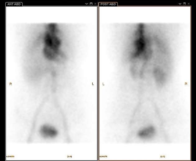
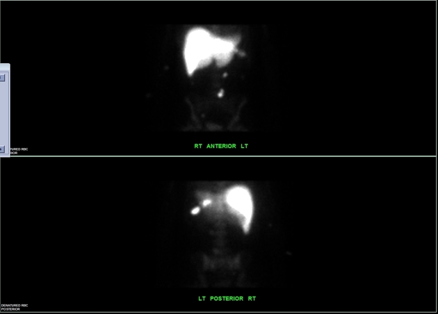
Scintigraphy performed with Tc-99m labeled RBCs
Citation, DOI, disclosures and article data
At the time the article was created The Radswiki had no recorded disclosures.
View The Radswiki's current disclosuresAt the time the article was last revised Mostafa Elfeky had no financial relationships to ineligible companies to disclose.
View Mostafa Elfeky's current disclosures- Technetium-99m labeled red blood cell
Tc-99m labeled RBCs - with radiolabelling technique in vivo or in vitro of red cells 3 - is one of the technetium radiopharmaceuticals used in the non-invasive assessment of gastrointestinal (GI) bleeding 2, characterized by high sensitivity (93%) and specificity (95%) 4. It is capable, in fact, of targeting intestinal hemorrhages of a few milliliters immediately after the injection of the radioactive tracer or in the following 12-24 hours (diluition principle) 5,6.
There are three ways to label erythrocytes, namely in vivo, modified in vivo, and in vitro methods. In vivo method injects stannous pyrophosphate into the patient's blood, followed by Tc99m-pertechnetate injection. However, this method has low labeling efficiency. In the modified in vivo method, the blood is withdrawn after injecting the stannous pyrophosphate. The blood is then withdrawn from the patient mixed with Tc-99m-pertechnetate and injected back into the same patient. In the in vitro method, the blood is first withdrawn from the patient and mixed with stannous pyrophosphate and Tc-99m-pertechnetate using a cold kit. After mixing, the blood is then injected back into the patient. This method has the highest labeling efficiency 7.
Stannous pyrophosphate is important because it acts as a reducing agent in order to prevent pertechnetate ions from diffusing into the extravascular spaces, affecting the labeling efficacy of the RBCs 8.
Characteristics
photon energy: 140 KeV
physical half-life: 6 hours
biological half-life:
normal distribution: heart, vessels, spleen
-
miscellaneous facts:
threshold for detection is 0.05-0.1 mL/min
angiography threshold is 0.5-1 mL/min
the criteria for positive scan are qualitative type: hot spot appears and conforms to intestinal anatomy, abnormal activity increases over time, activity moves antegrade or retrograde through bowel
Uses, doses and timings
timing: immediate/angioscintigraphic phase (1 sec frames x 60 sec); 1 min frames x 60-90 min; delayed imaging if necessary (6-8-24 hrs).

References
- 1. Emslie JT, Zarnegar K, Siegel ME, Beart RW. Technetium-99m-labeled red blood cell scans in the investigation of gastrointestinal bleeding. (1996) Diseases of the colon and rectum. 39 (7): 750-4. Pubmed
- 2. Grady E, Grady. Gastrointestinal Bleeding Scintigraphy in the Early 21st Century. (2016) Journal of nuclear medicine : official publication, Society of Nuclear Medicine. doi:10.2967/jnumed.115.157289 - Pubmed
- 3. Srivastava S, Srivastava CL, Srivastava. Radionuclide-labeled red blood cells: current status and future prospects. (1984) Seminars in nuclear medicine. doi:10.1016/s0001-2998(84)80022-7 - Pubmed
- 4. Bunker S, Bunker LR, Bunker TD, Bunker RM, Bunker RJ, Bunker BJ, Bunker BM, Bunker MR, Bunker RL, Bunker LA, Bunker. Scintigraphy of gastrointestinal hemorrhage: superiority of 99mTc red blood cells over 99mTc sulfur colloid. (1984) AJR. American journal of roentgenology. doi:10.2214/ajr.143.3.543 - Pubmed
- 5. Winzelberg G, Winzelberg MK, Winzelberg SH, Winzelberg WA, Winzelberg GA, Winzelberg. Evaluation of gastrointestinal bleeding by red blood cells labeled in vivo with technetium-99m. (1979) Journal of nuclear medicine : official publication, Society of Nuclear Medicine. doi: - Pubmed
- 6. Zuckerman D, Zuckerman BT, Zuckerman BE, Zuckerman. Massive hemorrhage in the lower gastrointestinal tract in adults: diagnostic imaging and intervention. (1993) AJR. American journal of roentgenology. doi:10.2214/ajr.161.4.8372742 - Pubmed
- 7. Grady E. Gastrointestinal Bleeding Scintigraphy in the Early 21st Century. J Nucl Med. 2015;57(2):252-9. doi:10.2967/jnumed.115.157289 - Pubmed
- 8. Front D, Israel O, Groshar D, Weininger J. Technetium-99m-Labeled Red Blood Cell Imaging. Semin Nucl Med. 1984;14(3):226-50. doi:10.1016/s0001-2998(84)80017-3
Incoming Links
Related articles: Imaging technology
- imaging technology
- imaging physics
- imaging in practice
-
x-rays
- x-ray physics
- x-ray in practice
- x-ray production
- x-ray tube
- filters
- automatic exposure control (AEC)
- beam collimators
- grids
- air gap technique
- cassette
- intensifying screen
- x-ray film
- image intensifier
- digital radiography
- digital image
- mammography
- x-ray artifacts
- radiation units
- radiation safety
- radiation detectors
- fluoroscopy
-
computed tomography (CT)
- CT physics
- CT in practice
- CT technology
- CT image reconstruction
- CT image quality
- CT dose
-
CT contrast media
-
iodinated contrast media
- agents
- water soluble
- water insoluble
- vicarious contrast material excretion
- iodinated contrast media adverse reactions
- agents
- non-iodinated contrast media
-
iodinated contrast media
-
CT artifacts
- patient-based artifacts
- physics-based artifacts
- hardware-based artifacts
- ring artifact
- tube arcing
- out of field artifact
- air bubble artifact
- helical and multichannel artifacts
- CT safety
- history of CT
-
MRI
- MRI physics
- MRI in practice
- MRI hardware
- signal processing
-
MRI pulse sequences (basics | abbreviations | parameters)
- T1 weighted image
- T2 weighted image
- proton density weighted image
- chemical exchange saturation transfer
- CSF flow studies
- diffusion weighted imaging (DWI)
- echo-planar pulse sequences
- fat-suppressed imaging sequences
- gradient echo sequences
- inversion recovery sequences
- metal artifact reduction sequence (MARS)
-
perfusion-weighted imaging
- techniques
- derived values
- saturation recovery sequences
- spin echo sequences
- spiral pulse sequences
- susceptibility-weighted imaging (SWI)
- T1 rho
- MR angiography (and venography)
-
MR spectroscopy (MRS)
- 2-hydroxyglutarate peak: resonates at 2.25 ppm
- alanine peak: resonates at 1.48 ppm
- choline peak: resonates at 3.2 ppm
- citrate peak: resonates at 2.6 ppm
- creatine peak: resonates at 3.0 ppm
- functional MRI (fMRI)
- gamma-aminobutyric acid (GABA) peak: resonates at 2.2-2.4 ppm
- glutamine-glutamate peak: resonates at 2.2-2.4 ppm
- Hunter's angle
- lactate peak: resonates at 1.3 ppm
- lipids peak: resonates at 1.3 ppm
- myoinositol peak: resonates at 3.5 ppm
- MR fingerprinting
- N-acetylaspartate (NAA) peak: resonates at 2.0 ppm
- propylene glycol peak: resonates at 1.13 ppm
-
MRI artifacts
- MRI hardware and room shielding
- MRI software
- patient and physiologic motion
- tissue heterogeneity and foreign bodies
- Fourier transform and Nyquist sampling theorem
- MRI contrast agents
- MRI safety
-
ultrasound
- ultrasound physics
-
transducers
- linear array
- convex array
- phased array
- frame averaging (frame persistence)
- ultrasound image resolution
- imaging modes and display
- pulse-echo imaging
- real-time imaging
-
Doppler imaging
- Doppler effect
- color Doppler
- power Doppler
- B flow
- color box
- Doppler angle
- pulse repetition frequency and scale
- wall filter
- color write priority
- packet size (dwell time)
- peak systolic velocity
- end-diastolic velocity
- resistive index
- pulsatility index
- Reynolds number
- panoramic imaging
- compound imaging
- harmonic imaging
- elastography
- scanning modes
- 2D ultrasound
- 3D ultrasound
- 4D ultrasound
- M-mode
-
ultrasound artifacts
- acoustic shadowing
- acoustic enhancement
- beam width artifact
- reverberation artifact
- ring down artifact
- mirror image artifact
- side lobe artifact
- speckle artifact
- speed displacement artifact
- refraction artifact
- multipath artifact
- anisotropy
- electrical interference artifact
- hardware-related artifacts
- Doppler artifacts
- aliasing
- tissue vibration
- spectral broadening
- blooming
- motion (flash) artifact
- twinkling artifact
- acoustic streaming
- biological effects of ultrasound
- history of ultrasound
-
nuclear medicine
- nuclear medicine physics
- detectors
- tissue to background ratio
-
radiopharmaceuticals
- fundamentals of radiopharmaceuticals
- radiopharmaceutical labeling
- radiopharmaceutical production
- nuclear reactor produced radionuclides
- cyclotron produced radionuclides
- radiation detection
- dosimetry
- specific agents
- carbon-11
- chromium-51
- fluorine agents
- gallium agents
- Ga-67 citrate
- Ga-68
- iodine agents
-
I-123
- I-123 iodide
- I-123 ioflupane (DaTSCAN)
- I-123 ortho-iodohippurate
- I-131
-
MIBG scans
- I-123 MIBG
- I-131 MIBG
-
I-123
- indium agents
- In-111 Octreoscan
- In-111 OncoScint
- In-111 Prostascint
- In-111 oxine labeled WBC
- krypton-81m
- nitrogen-13
- oxygen-15
- phosphorus-32
- selenium-75
-
technetium agents
- Tc-99m DMSA
- Tc-99m DTPA
- Tc-99m DTPA aerosol
- Tc-99m HMPAO
- Tc-99m HMPAO labeled WBC
- Tc-99m MAA
- Tc-99m MAG3
- Tc-99m MDP
- Tc-99m mercaptoacetyltriglycine
- Tc-99m pertechnetate
- Tc-99m labeled RBC
- Tc-99m sestamibi
- Tc-99m sulfur colloid
- Tc-99m sulfur colloid (oral)
- thallium-201 chloride
- xenon agents
- in vivo therapeutic agents
- pharmaceuticals used in nuclear medicine
-
emerging methods in medical imaging
- radiography
- phase-contrast imaging
- CT
- deep-learning reconstruction
- photon counting CT
- virtual non-contrast imaging
- ultrasound
- magnetomotive ultrasound (MMUS)
- superb microvascular imaging
- ultrafast Doppler imaging
- ultrasound localization microscopy
- MRI
- nuclear medicine
- total body PET system
- immuno-PET
- miscellaneous
- radiography


 Unable to process the form. Check for errors and try again.