
Thoracic aortic aneurysms are a type of thoraco-abdominal aneurysm. They are much less common than abdominal aortic aneurysms (0.16% vs 4%) 17. There is a wide range of causes, and the ascending aorta is the segment most commonly affected. Both CT-angiography and MR-angiography are the modalities of choice to image this condition.
On this page:
Terminology
The normal aortic diameter varies based on age, sex, and body surface area. In general, the term aneurysm is used when the axial diameter is >5.0 cm for the ascending aorta and >4.0 cm for the descending aorta 12. When enlarged above normal but not reaching aneurysmal definition, the terms dilatation/ectasia can be used 9,12.
Epidemiology
Most commonly occur in the 50 to 60-year-old age group with an estimated incidence of ~7.5 per 100,000 patient-years 8. There is a male predominance (M:F is 3:1).
Clinical presentation
Thoracic aneurysms are often incidental findings on chest imaging. They may present symptomatically if large enough to cause mass effect on the airway, esophagus or pulmonary vasculature. Alternatively, they may present due to a complication, including rupture, dissection, aorto-bronchial or aorto-esophageal fistulae.
Pseudoaneurysms of the thoracic aorta are usually the result of significant thoracic trauma, both penetrating and blunt, and carry a very high mortality, with 80-90% of patients dying before reaching hospital 4.
Pathology
Location
Aneurysmal dilatation can affect any part of the thoracic aorta. Relative frequencies are (with some involving more than one segment) 7:
aortic root/ascending aorta: 60%
aortic arch: 10% - aortic arch aneurysms
descending aorta: 40%
thoracoabdominal segment: 10%
Etiology
Thoracic aortic aneurysms can be divided pathologically according to their relationship to the aortic wall 1:
-
atherosclerotic aneurysms (most common)
-
inflammatory/aortitis
-
connective tissue disease
-
cystic medial necrosis
-
-
ciprofloxacin use (maybe quinolone class effect) 13
quinolones promote loss of extracellular structural integrity, by several non-antimicrobial mechanisms 13
in the UK, caution is now advised in using quinolones in high-risk patients 14
-
trauma
post-surgery
Associations
intracranial cerebral aneurysms (~10% prevalence) 11
Radiographic features
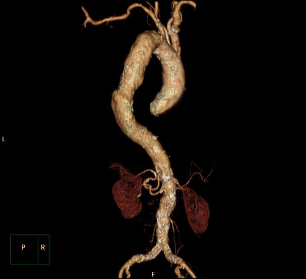
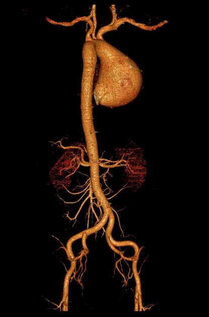
The location and shape of thoracic aortic aneurysms are variable. An aortic aneurysm, as aneurysms elsewhere, can be described as saccular or fusiform. In the case of fusiform dilatation, the term aneurysm should be applied when the diameter is >4 cm 1.
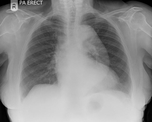
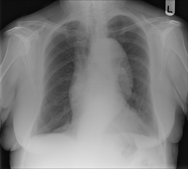
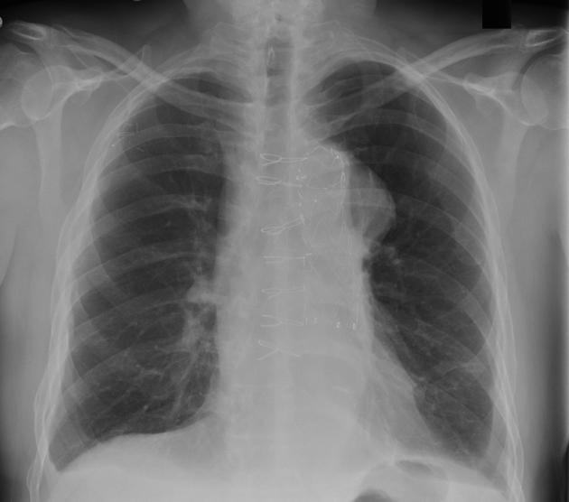
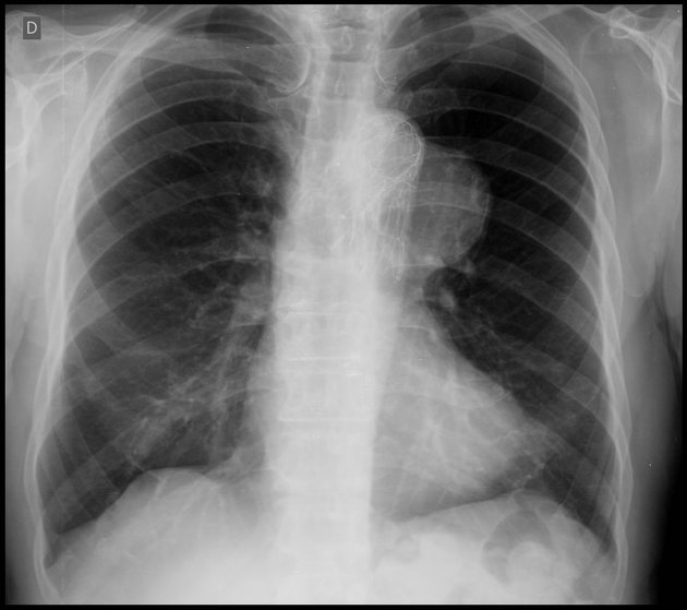
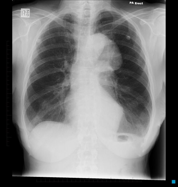
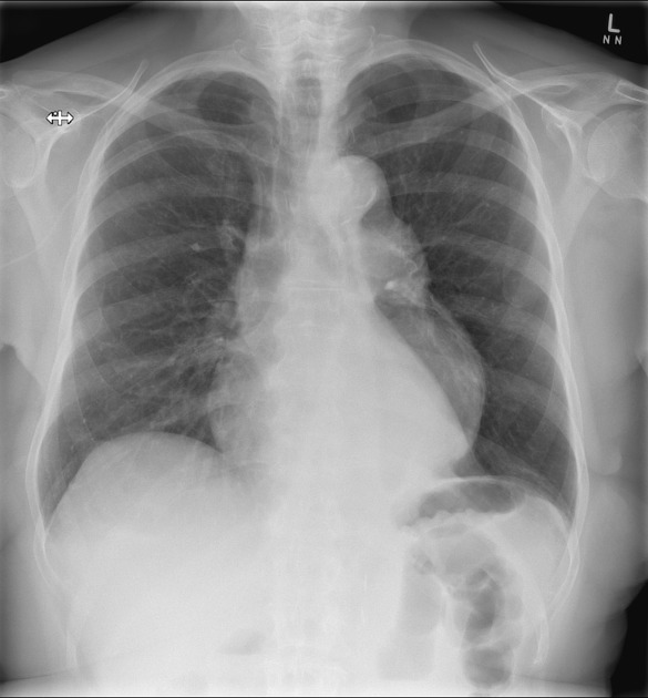
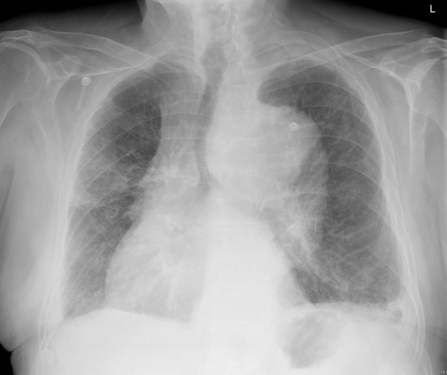
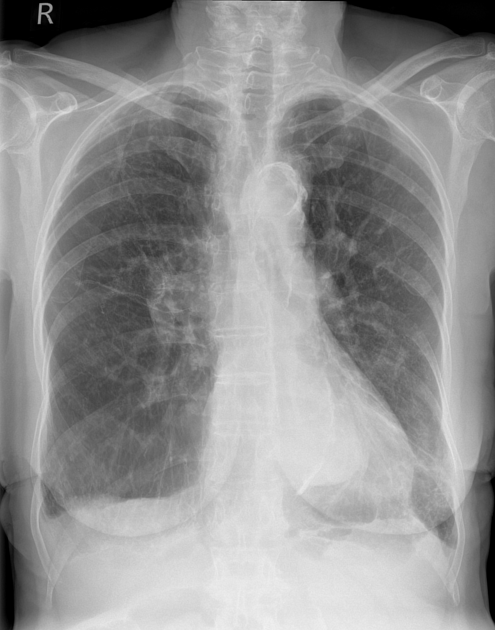
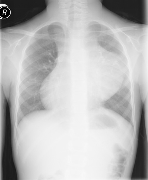
Plain radiograph
The thoracic aorta can usually be seen on both frontal and lateral chest radiographs, and aneurysms are often obvious. However, it is difficult to assess size accurately (due to magnification effects and often poor visualization on the side of the artery).
Additionally, mediastinal masses may mimic aortic aneurysms.
Mural calcification is seen both in atherosclerotic disease as well as various causes of aortitis (see causes of ascending aorta calcification).
Ultrasound
Unlike abdominal aneurysms that can usually be readily assessed and monitored with ultrasound, thoracic aortic aneurysms are encased in bone and air making transthoracic ultrasound unsuitable.
Transesophageal echocardiography (TOE) can visualize much of the descending aorta, but due to its invasive nature is not routinely used.
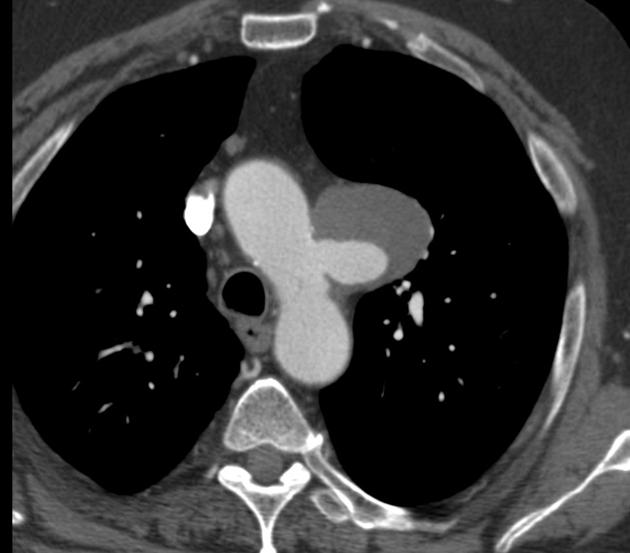
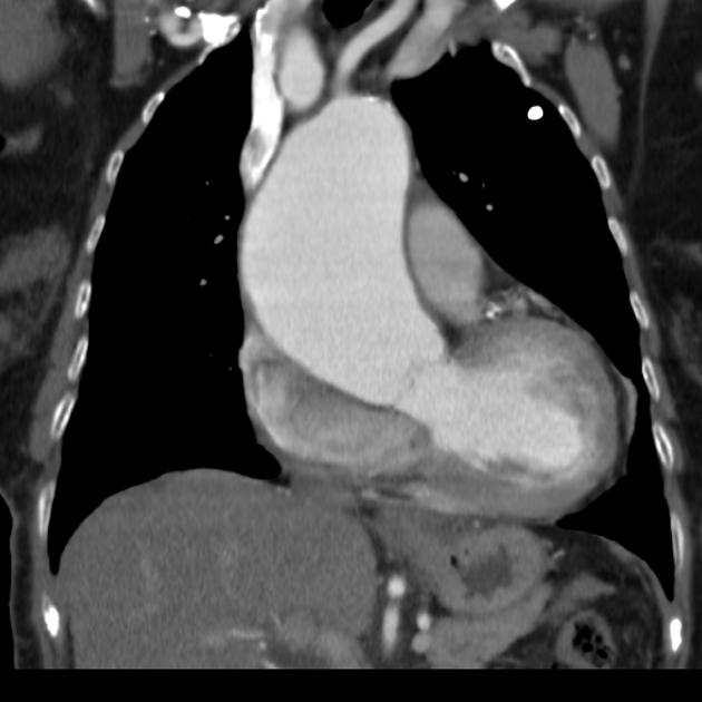
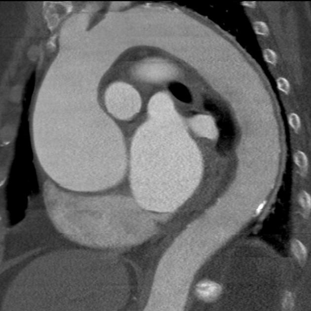
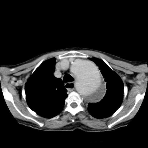
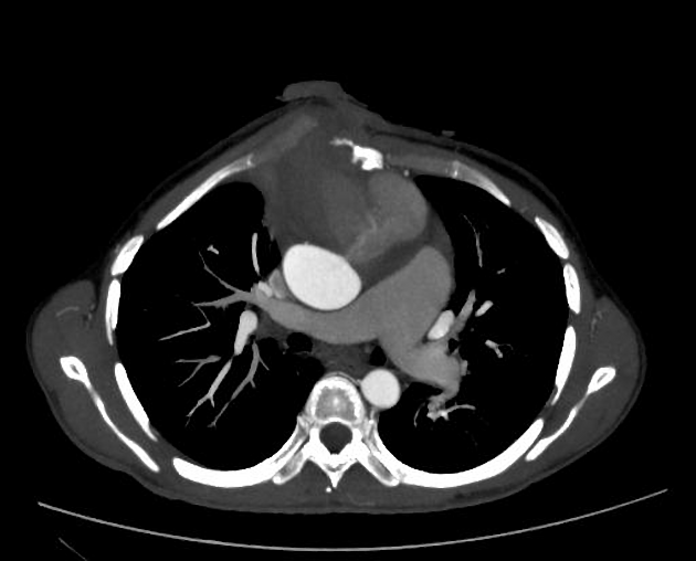
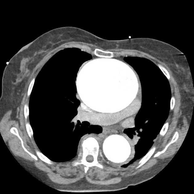
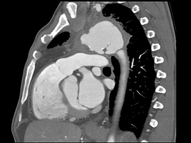
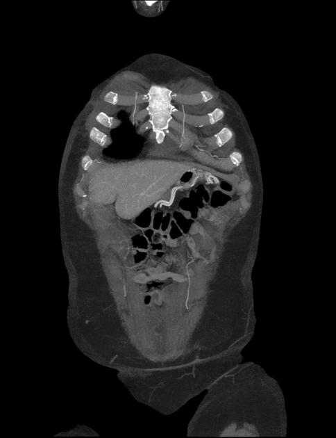
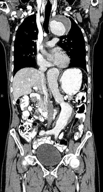
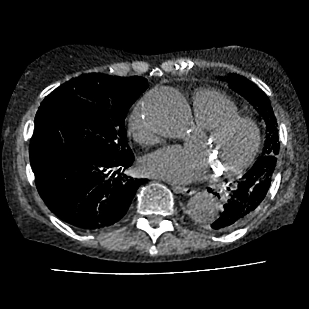
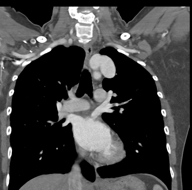
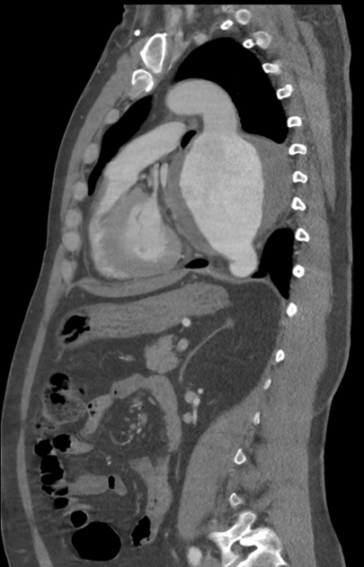
CT
CTA is the work-horse of aneurysm assessment and is able to rapidly image the relevant vascular territory with high resolution. It can visualize both the sac and the lumen and detect potential complications.
Typically, aneurysms appear as dilatations of the lumen. The walls may be thin or thickened by the presence of a mural thrombus (circumferential or more frequently eccentric).
Calcified atherosclerotic disease is often identified not only in the wall of an aneurysm but adjacent arteries.
If rupture or leak has occurred hematoma/fluid may be seen adjacent to the aorta, in the left pleural cavity or the pericardium 1.
See main article: reporting tips for aortic aneurysms.
Angiography (DSA)
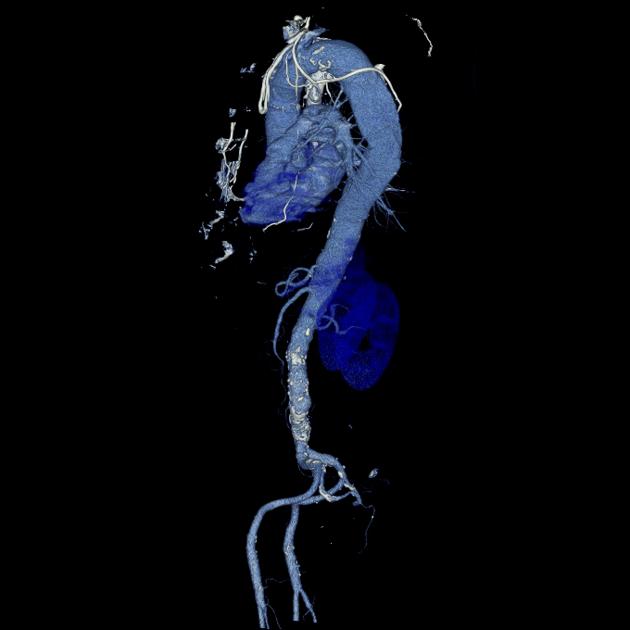
Although angiography has long been considered the gold standard for vascular imaging, it has largely been superseded by CTA and MRA, which can obtain 3D volumetric data, and able to assess the extraluminal soft tissues.
Angiography is however used during endovascular repair.
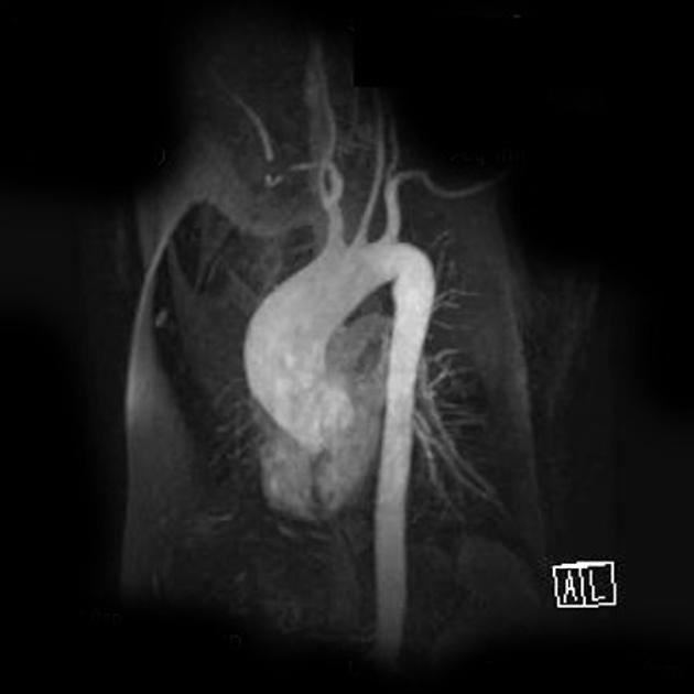
MRI
MRI has the advantage of not requiring ionizing radiation or large volumes of iodinated contrast 2. This is particularly advantageous in young patients with connective tissue disorders. However, there are limitations in patients with pacemakers and those with reduced renal function (see nephrogenic systemic fibrosis).
Acquisitions capable of being reformatted in three dimensions are essential to allow for accurate luminal measurement 2.
Treatment and prognosis
Mild to moderate aneurysmal dilatation can usually be treated conservatively and monitored. When the diameter reaches 5-6 cm intervention is usually considered as the risk of rupture is significantly elevated 1. Treatment options include:
-
open repair
In general, when possible, endovascular repair (TEVAR) is the treatment of choice, with reduced morbidity and mortality 2.
The majority of patients with thoracic aortic aneurysms either die of a direct complication of the aneurysm (i.e. rupture) or other cardiovascular complications 3. The main predictors of rupture are the size and speed of growth 3. The average growth of a thoracic aneurysm appears to be lower than that of abdominal aneurysms, typically in the order of 1-2 mm/year, and correlates with a better prognosis for thoracic aneurysms when controlled for size 3.
Complications
rupture
distal embolization
-
fistula formation
Differential diagnosis
aortic spindle: circumferential bulge at the proximal descending thoracic aorta just beyond the aortic isthmus (normal anatomical variant)


 Unable to process the form. Check for errors and try again.