
Warthin tumors, also known as lymphomatous papillary cystadenomas, are benign, sharply demarcated salivary gland tumors, almost exclusively encountered in the parotid gland. They are of lymphoid origin and may be bilateral or multifocal in up to 20% of cases and are the most common neoplastic cause of multiple solid parotid masses.
On this page:
Epidemiology
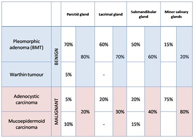
Warthin tumors are the 2nd most common benign parotid gland tumor (after pleomorphic adenoma) and represent up to 10% of all parotid tumors. They are the most common bilateral or multifocal benign parotid tumors. They typically occur in older age (6th decade) and are twice as common in men (2.2:1) 11.
Associations
smoking
irradiation
Clinical presentation
Patients typically present with painless parotid swelling.
Pathology
Location
They tend to favor the parotid tail region at the level of the mandibular angle. In <10% cases, they are found elsewhere, including the submandibular glands, cervical lymph nodes, and ectopic nests of salivary tissue, e.g. larynx, maxillary antrum, oral cavity (e.g. lower lip, buccal mucosa) 11,12.
Radiographic features
They are often multicentric (20%) and are usually small (1-4 cm). They have a typically heterogeneous appearance on all modalities, often with cystic components (30%). The tendency to undergo cystic change is greater than any other salivary gland tumor 4,5.
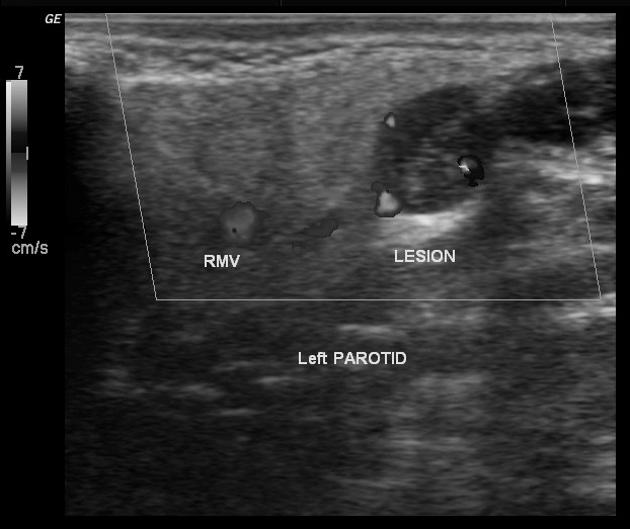
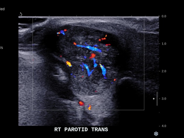
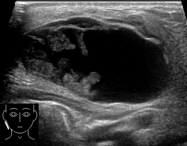
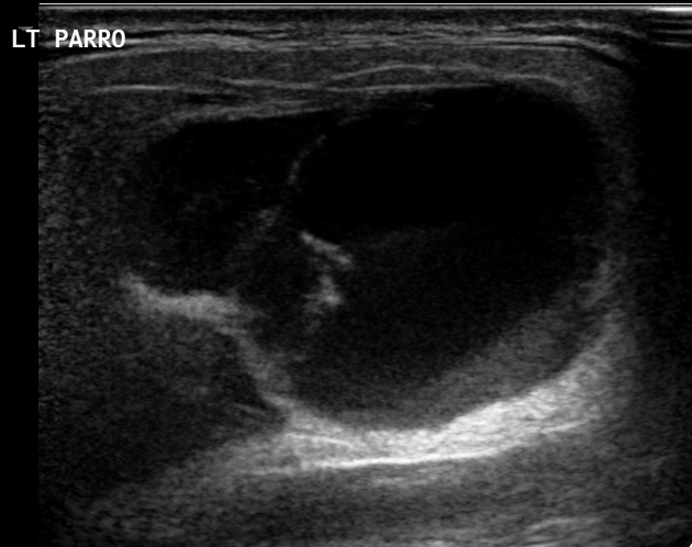
Ultrasound
Most tumors tend to be ovoid, with well-defined margins and multiple irregular, small, sponge-like anechoic areas 10. Tumors that are large (e.g. >5 cm) tend to have a higher proportion of cystic content than smaller lesions had and in some cases can be composed almost entirely of cystic material. They are often hypervascular.
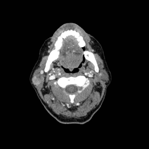
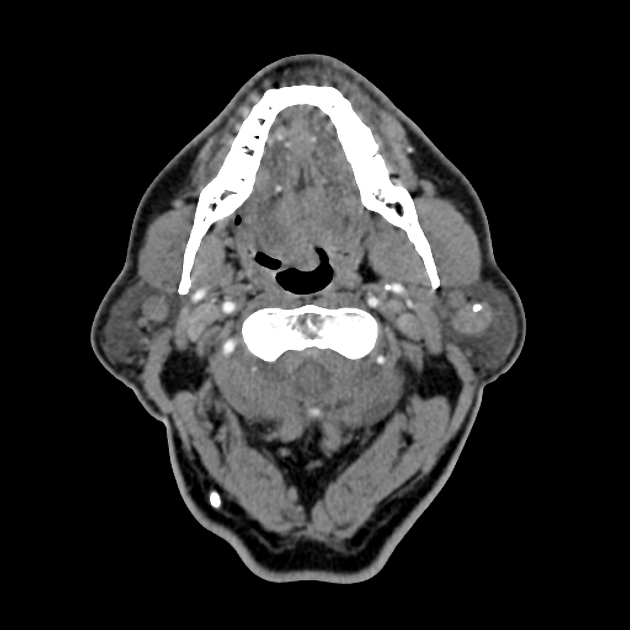
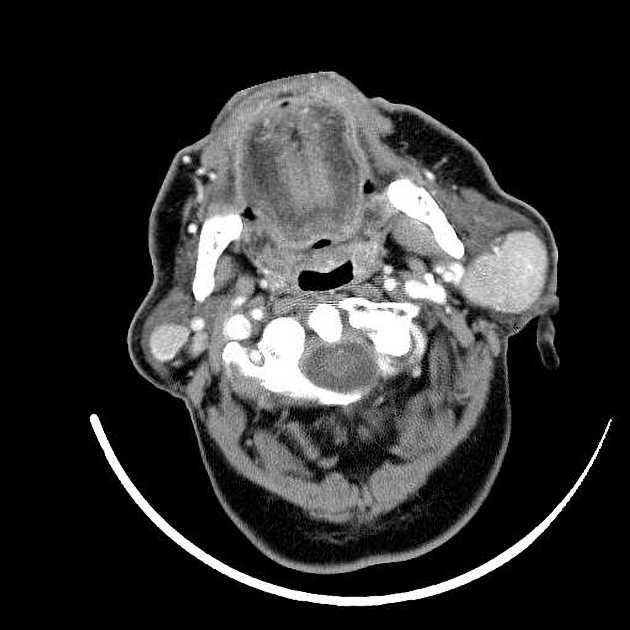
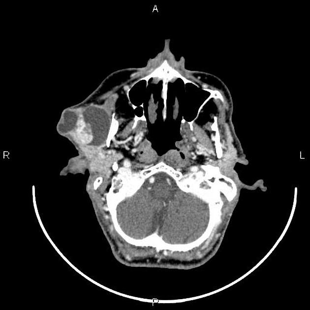
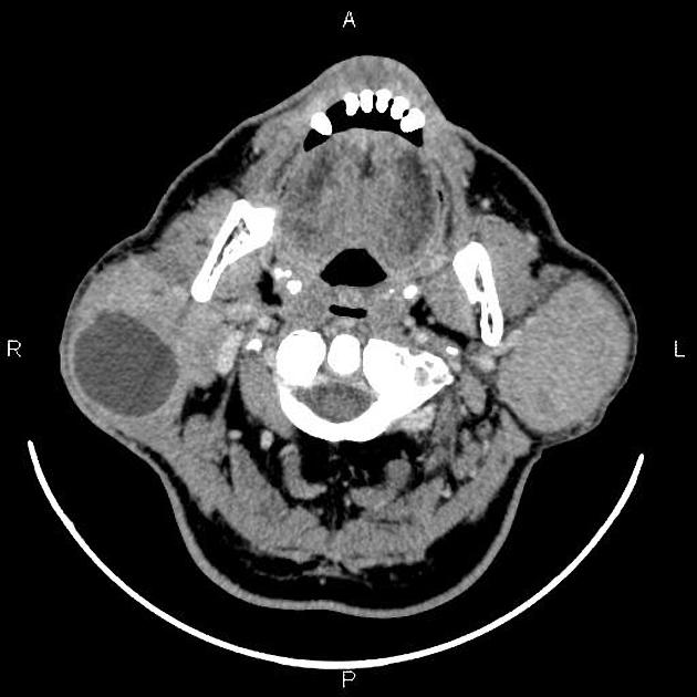
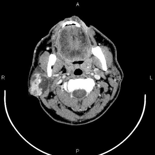
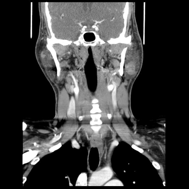
CT
classic appearance is a well-defined heterogeneous solid cystic lesion within the superficial lobe of parotid/parotid tail
well defined
no calcification
cystic changes appear as intralesional lower attenuation
moderate enhancement
presence of a mural nodule is strongly suggestive of Warthin tumor
can be often seen bilaterally
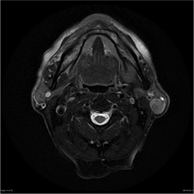
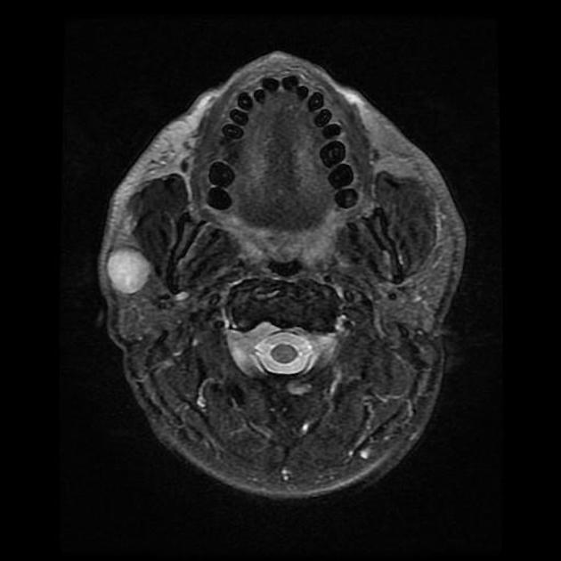
MRI
Signal characteristics
T1: low to intermediate signal with cyst containing cholesterol components containing focal high signal 2
T2: heterogeneous and variable signal intensity
T1 C+ (Gd): cystic components do not take up contrast while solid parts usually enhance 1
Nuclear medicine
Warthin tumors often shows uptake with Tc99-pertechnetate, thallium, and FDG-PET 7.
Treatment and prognosis
They are benign with an extremely low incidence of malignant transformation (~1%). Some advocate surgical excision while others favor conservative management with follow-up imaging. The commonest surgery is a superficial parotidectomy and recurrence rate is low, less than 5% in one of the largest published series 11,12.
History and etymology
These lesions were first described in 1929 by an American pathologist, Aldred Scott Warthin 13,14.
Differential diagnosis
Possible imaging differential considerations include:
-
other salivary gland tumors
acinic cell carcinoma (of salivary gland)
infiltrative lesion, e.g. sarcoidosis


 Unable to process the form. Check for errors and try again.