
Presentation
Assessment of liver disease.
Patient Data


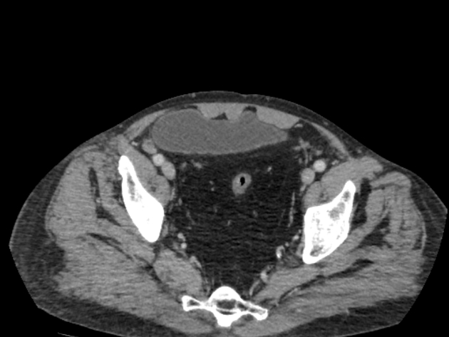
Marked pelvic lipomatosis with displacement of the urinary bladder anteriorly, stretched rectum and sigmoid as well as displacement of bowel loops peripherally.
Also noted: liver cirrhosis with periportal fibrosis, bilateral simple renal cysts, bilateral hydrocele marked on left side, splenectomy, small epigastric hernia transmitting omental fat and gall bladder stone disease.
Case Discussion
Pelvic lipomatosis represents excessive deposition of fat in pelvis leading to compression of pelvic organs. Patients usually present with dysuria, urgency, urinary incontinence, constipation and can be asymptomatic. Imaging features include inverted teardrop-shaped bladder (pear-shaped bladder), elongated rectum that is symmetrically compressed and peripheral displacement of bowel loops.


 Unable to process the form. Check for errors and try again.