
Presentation
Chronic abdominal pain of about 2 years duration, occasional vomiting, constipation and anemia
Patient Data
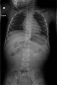
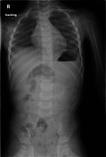

Abdominal radiograph shows soft tissue mass with central calcifications in the left hemiabdomen adjacent to L3 vertebra.
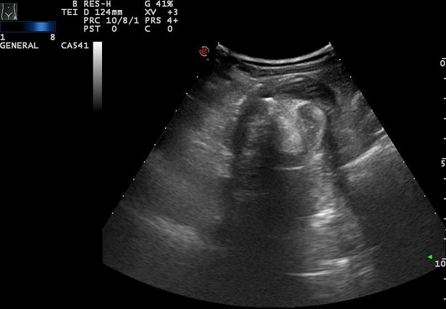
A mixed echogenic (hyperechoic with distant acoustic shadowing and hypoechoic) mass is noted in the left upper abdomen, anterior to the left kidney. It measures 55 x 63 x 40 mm. Abdominal CT was requested to further characterize the lesion.


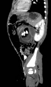
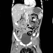
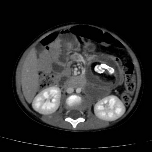
There is a large mixed density mass, related to proximal jejunal bowel loops mesentery in the left upper abdomen, anterior to the left kidney. The mass measures about 5 x 6 x 4 cm and shows fatty density with multiple intralesional calcifications (teeth \ bones). The appearance is in keeping with mesenteric teratoma.
Case Discussion
Even though teratomas are usually asymptomatic, they sometimes present with symptoms due to local compression as with this case where the child presented with chronic abdominal pain, occasional vomiting, constipation and refusal of feeds which led to anemia on two occasions. This prompted further investigations and the characteristic presence of tooth-like structures, bones and fat components are characteristic for teratomas. However, child was referred to a higher center for surgery and histopathology confirmation.


 Unable to process the form. Check for errors and try again.