
Patient Data
Note: This case has been tagged as "legacy" as it no longer meets image preparation and/or other case publication guidelines.
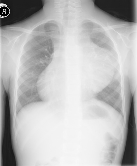
There is a large anterior mediastinal mass measuring 9 cm in size with calcification within it. There is shift of the mediastinum towards right side. No pleural effusion is seen. Findings are most likely suggestive of teratoma.
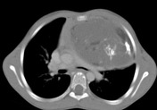


Anterior hypodense well defined left-sided mediastinal mass, with calcification and fat within it, the appearance is highly suggestive of teratoma.
Case Discussion
Frontal chest radiograph demonstrates a left paratracheal radio-opacity with sharp lateral margin and indistinct medial border that silhouette the left cardiac border, suggestive of anterior mediastinal mass. Shift of the mediastinum to the right side is also seen. Specks of calcification within the mass is noted. Findings are most likely suggestive of teratoma.
Selected axial and sagittal chest CT confirm the presence of anterior hypodense well defined left-sided mediastinal mass, with calcification and fat within it, the appearance is highly suggestive of teratoma.


 Unable to process the form. Check for errors and try again.