
Note: This case has been tagged as "legacy" as it no longer meets image preparation and/or other case publication guidelines.

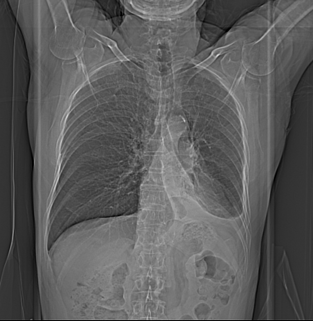
CT scoutogram demonstrates bulkiness of the left hilum with collapse of the left lower lobe and a small pleural effusion.
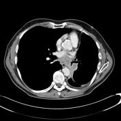
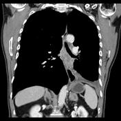
CT through the chest with contrast demonstrates numerous necrotic lymph nodes in the subcarinal region and left hilum with almost complete obliteration of left lower lobe bronchus with left lower lobe collapse.
There is a tiny left pleural effusion and bilateral enhancing adrenal lesions.
Enhancing right adrenal mass.
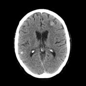
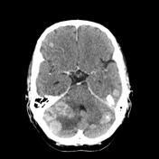
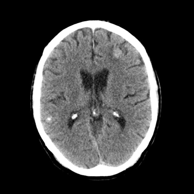
Multiple enhancing nodules scattered throughout cerebral and cerebellar hemispheres bilaterally are in keeping with cerebral metastases, some of which are hemorrhagic.
Case Discussion
Findings are consistent with the subsequently pathology proven bronchogenic small cell carcinoma.


 Unable to process the form. Check for errors and try again.