
Presentation
Work up for hematuria.
Patient Data


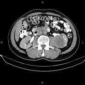

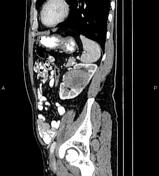
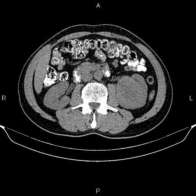
A 68 x 55 mm well-defined mass is seen in the mid portion of the left kidney that bulges into the urinary collecting system. The neoplasm has a similar density to normal renal parenchyma on non-enhanced CT scan. After IV contrast media injection, the attenuation value increased from 28 HU to 47 HU. Nephrographic-phase CT scan shows that the mass enhances less than normal renal parenchyma. There is no sign of local invasion, no vascular extension, and no regional lymphadenopathy.
A 7 mm cyst is present at the left liver lobe. In addition, an 11 mm hyper vascular mass with persistent enhancement is noted at the right liver lobe, most consistent with flash type hemangioma.
The prostate gland is enlarged.
Case Discussion
The patient went on to have a left nephrectomy, and histopathology evaluation confirmed renal cell carcinoma.


 Unable to process the form. Check for errors and try again.