
Presentation
Neck and shoulder pain.
Patient Data
Note: This case has been tagged as "legacy" as it no longer meets image preparation and/or other case publication guidelines.
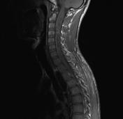
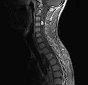
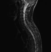
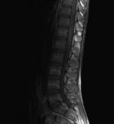
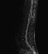
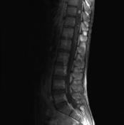

MRI spine reveals an enhancing intramedullary lesion in the cervical and lumbar spine associated with syringomyelia.
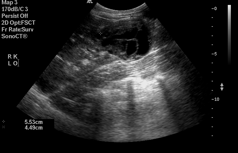
There are multiple cystic lesions of the pancreas and kidneys. Most of these cysts appear simple apart from right kidney lower pole cyst which appears more complex and suspicious for malignancy (follow-up unavailable).


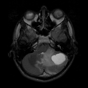
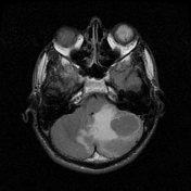
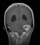
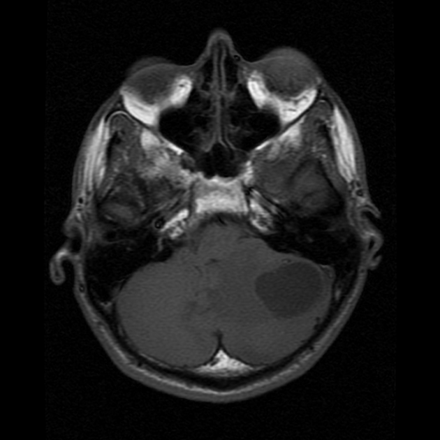
The patient presented 3 years later with a severe headache. MRI of the brain performed which reveals multiple enhancing cerebellar lesion the largest seen in the left cerebellum, probably corresponding to the small enhancing nodule seen 3 years earlier. Associated with acute hydrocephalus. Again demonstrated an enhancing lesion of the left eye globe with a new lesion of the right one probably represent retinal hemangioblastomas.
The lumbar lesion was surgically removed and pathologically proven hemangioblastoma.
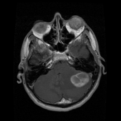
MRI brain was done for screening of posterior fossa hemangioblastomas, which revealed a single small enhancing lesion in the left cerebellar hemisphere. Enhancing lesion of the left eye globe likely represents retinal hemangioblastoma; features are therefore in keeping with von Hippel Lindau syndrome.
Case Discussion
Spinal hemangioblastomas are less common than those located in the cerebellar hemispheres (accounting for 80% of such tumors). They are commonly located on the posterior aspect of the spinal cord and 70% are associated with syringomyelia or cystic component. Contrast-enhanced MRI optimizes visualization of the small mural nodules.
The combination of multiple hemangioblastomas, retinal lesions and multiple renal and pancreatic cysts is characteristic of von Hippel Lindau disease.


 Unable to process the form. Check for errors and try again.