
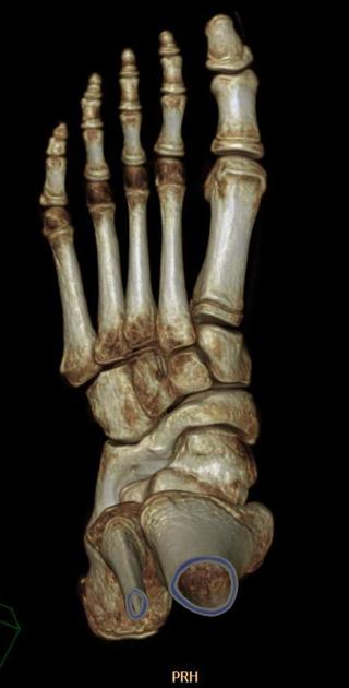
Calcaneonavicular coalition
Citation, DOI, disclosures and article data
At the time the article was created Alexandra Stanislavsky had no recorded disclosures.
View Alexandra Stanislavsky's current disclosuresAt the time the article was last revised Joachim Feger had no financial relationships to ineligible companies to disclose.
View Joachim Feger's current disclosures- Calcaneo-navicular coalition
Calcaneonavicular coalition is one of the two most common subtypes of the tarsal coalition, the other being talocalcaneal coalition. As with any coalition, it may be osseous (synostosis), cartilaginous (synchondrosis) or fibrous (syndesmosis).
Radiographic features
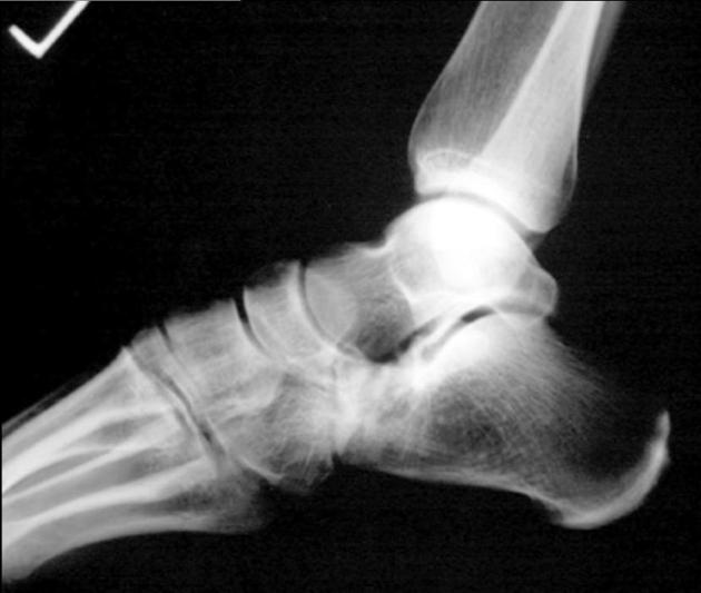
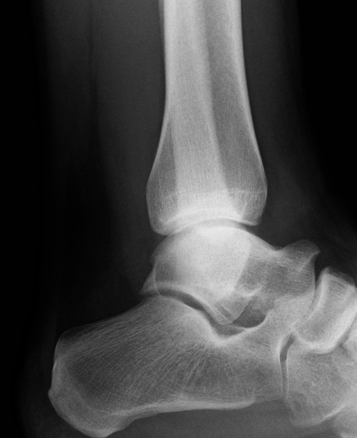
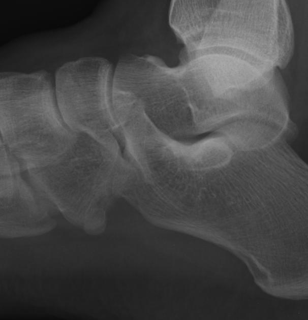
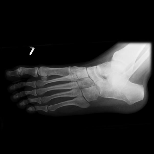
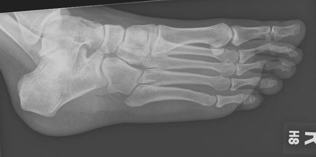
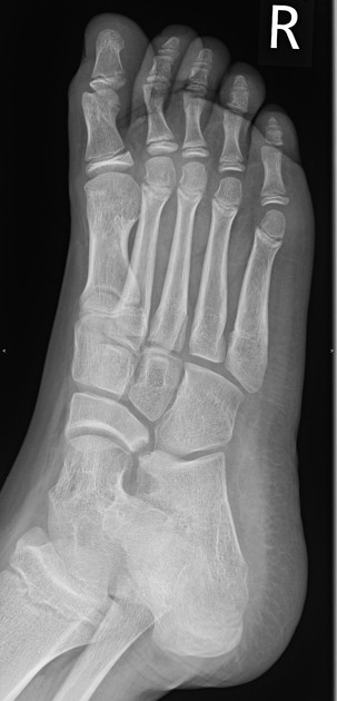
This type of coalition is more easily diagnosed on plain film than talocalcaneal coalition.
Plain radiograph
Oblique view
best at depicting calcaneonavicular coalition directly as a calcaneonavicular bar
AP view
may also directly show the coalition
-
indirect signs include
the broad proximal surface of the navicular bone: broader than the articulating talar head
lateral tapering of navicular
Lateral view
Indirect signs include:
anteater sign: an elongated anterior process of the calcaneus
reverse anteater sign: elongated lateral navicular
short talar neck
CT
CT can be used to confirm the diagnosis where this was equivocal or not seen on plain films. It may also be used for surgical planning.
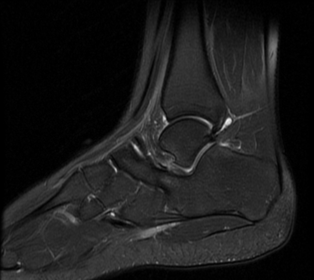
MRI
MRI is probably more helpful in assessing and characterizing cartilaginous and fibrous coalition and allows assessment of associated bone and soft tissue edema.
ADVERTISEMENT: Supporters see fewer/no ads
Treatment and prognosis
As with any tarsal coalition, non-operative management may allow some improvement in symptoms initially, but they usually return. Usually, surgical treatment with excision of the coalition is required.
References
- 1. Newman J & Newberg A. Congenital Tarsal Coalition: Multimodality Evaluation with Emphasis on CT and MR Imaging. Radiographics. 2000;20(2):321-32; quiz 526. doi:10.1148/radiographics.20.2.g00mc03321 - Pubmed
- 2. Crim J & Kjeldsberg K. Radiographic Diagnosis of Tarsal Coalition. AJR Am J Roentgenol. 2004;182(2):323-8. doi:10.2214/ajr.182.2.1820323 - Pubmed
- 3. Nalaboff K & Schweitzer M. MRI of Tarsal Coalition: Frequency, Distribution, and Innovative Signs. Bull NYU Hosp Jt Dis. 2008;66(1):14-21. - Pubmed
Incoming Links
- Calcaneonavicular coalition (bone scan)
- Talocalcaneal coalition
- Calcaneonavicular coalition
- Anteater nose
- Calcaneonavicular coalition
- Cuboid-navicular coalition
- Calcaneonavicular cartilage coalition
- Osteomyelitis and calcaneonavicular coalition
- Calcaneonavicular coalition
- Calcaneonavicular coalition - osseous
- Calcaneonavicular coalition
- Calcaneonavicular coalition
- Calcaneo-navicular coalition
- Calcaneonavicular coalition - anteater nose sign
- Calcaneonavicular coalition - cartilaginous
- Tarsal coalition
- Calcaneonavicular coalition - bilateral osseous
- Calcaneonavicular coalition
- Calcaneonavicular coalition
Related articles: Anatomy: Lower limb
- skeleton of the lower limb
- joints of the lower limb
-
hip joint
- ligaments
- muscles
- additional structures
- hip joint capsule
- zona orbicularis
- iliotibial band
-
hip bursae
- anterior
- iliopsoas bursa (iliopectineal bursa)
- lateral
- subgluteal bursae
- greater trochanteric bursa (subgluteus maximus bursa)
- subgluteus medius bursa
- subgluteus minimus bursa
- gluteofemoral bursa
- subgluteal bursae
- postero-inferior
- anterior
- ossification centers
-
knee joint
- ligaments
- anterior cruciate ligament
- posterior cruciate ligament
- medial collateral ligament
- lateral collateral ligament
- meniscofemoral ligament (mnemonic)
-
posterolateral ligamentous complex
- arcuate ligament
- patellar tendon and quadriceps tendon
- anterolateral ligament
- posterior oblique ligament
- oblique popliteal ligament
- medial patellofemoral ligament
- additional structures
- extensor mechanism of the knee
- groove for the popliteus tendon
- knee bursae
- anterior bursae
- medial bursae
- lateral bursae
- posterior bursae
- knee capsule
- lateral patellar retinaculum
- medial patellar retinaculum
- menisci
- pes anserinus (mnemonic)
- ossification centers
- ligaments
- tibiofibular joints
-
ankle joint
- regional anatomy
- medial ankle
- lateral ankle
- anterior ankle
- ligaments
- medial collateral (deltoid) ligament
- lateral collateral ligament
- additional structures
- ankle bursae
- ossification centers of the ankle
- variants
- regional anatomy
- foot joints
- subtalar joint
- mid-tarsal (Chopart) joint
-
tarsometatarsal (Lisfranc) joint
- ligaments
- intermetatarsal joint
- metatarsophalangeal joint
- interphalangeal joint
- ossification centers
-
hip joint
- spaces of the lower limb
-
muscles of the lower limb
- muscles of the pelvic group
- muscles of the thigh
- muscles of the leg
- anterior compartment of the leg
- posterior compartments of the leg
- lateral compartment of the leg
- muscles of the foot
- dorsal muscles
- plantar muscles
- 1st layer
- 2nd layer
- 3rd layer
- 4th layer
- accessory muscles of the lower limb
- accessory gluteal muscles
-
accessory muscles of the ankle
- accessory peroneal muscles
- accessory flexor digitorum longus muscle
- accessory soleus muscle
- peroneocalcaneus internus muscle
- tibiocalcaneus internus muscle
- extensor hallucis capsularis tendon
- anterior fibulocalcaneus muscle
- accessory extensor digiti secundus muscle
- tibioastragalus anticus of Gruber muscle
- vascular supply of the lower limb
- arterial supply of the lower limb
- venous drainage of the lower limb
- innervation of the lower limb
- lymphatic system of the lower limb
- lymphatic pathways
- anteromedial group
- anterolateral group
- posteromedial group
- posterolateral group
- lower limb lymph nodes
- lymphatic pathways


 Unable to process the form. Check for errors and try again.