
Duodenal diverticula are outpouchings from the duodenal wall (intraluminal diverticulum discussed separately). They may result from mucosal prolapse or the prolapse of the entire duodenal wall and can be found at any point in the duodenum although are by far most commonly located along the medial wall of the second, or superior wall of the third, part of the duodenum.
Diverticula located at the ampulla of Vater may cause difficulty for endoscopists as they attempt to cannulate the biliary system.
On this page:
Clinical presentation
Duodenal diverticula are very common, found in up to 23% of asymptomatic patients 2, and in the vast majority remain asymptomatic throughout life. In 10% of patients, some symptoms are attributable to them, with only a minority requiring surgical intervention 2.
Pathology
There are two types of duodenal diverticula:
primary diverticulum
secondary diverticulum
A primary duodenal diverticulum occurs where there is a prolapse of mucosa through the muscularis propria. They usually occur within the 2nd part (62%) and less commonly in the 3rd (30%) and 4th (8%) parts. Unlike secondary diverticula they are rarely seen in the 1st part. When they occur in the 2nd part, most (88%) are seen on the medial wall around the ampulla, 8% are seen posteriorly and 4% on the lateral wall.
A secondary duodenal diverticulum results from prolapse of the entire duodenal wall and almost invariably occurs in the 1st part of the duodenum. These are true diverticula and are usually secondary to duodenal or periduodenal inflammation, such as from previous ulcer disease.
Location specific subtypes
Radiographic features
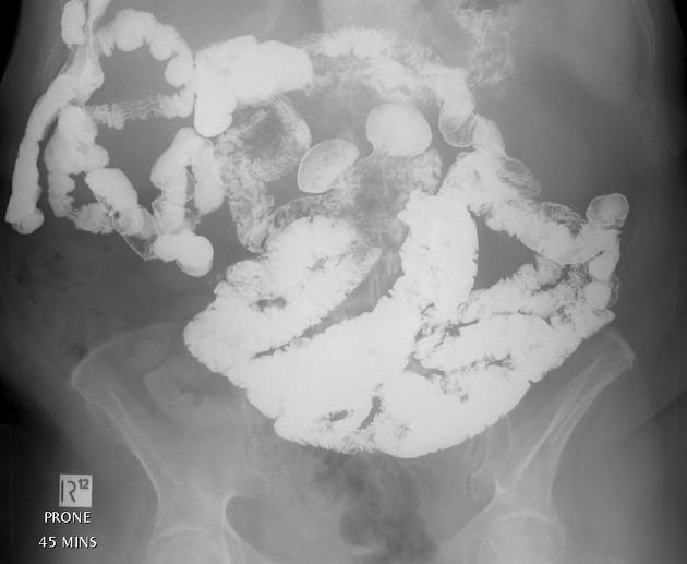
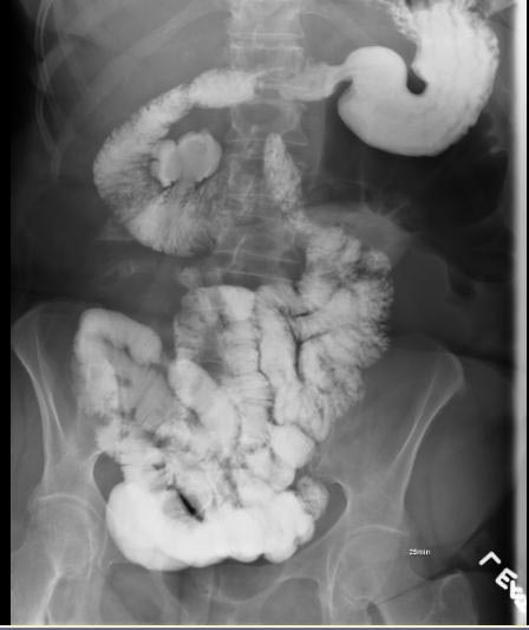
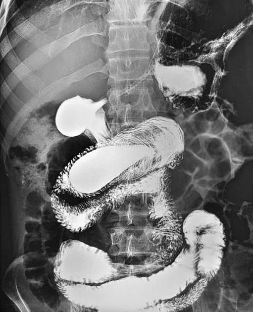
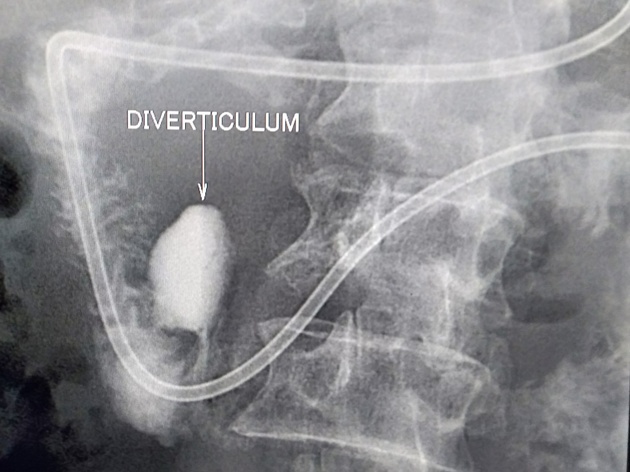
Fluoroscopy
Diverticula appear as extraluminal collection of barium contigous with the lumen, usually on the mesenteric side along the medial wall of D2 or the superior wall of D3. They can change configuration throughout the examination 7.
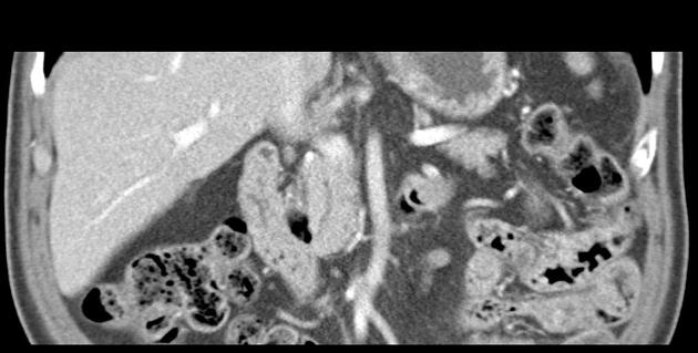
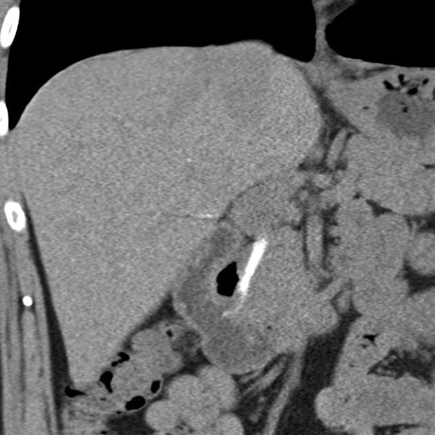
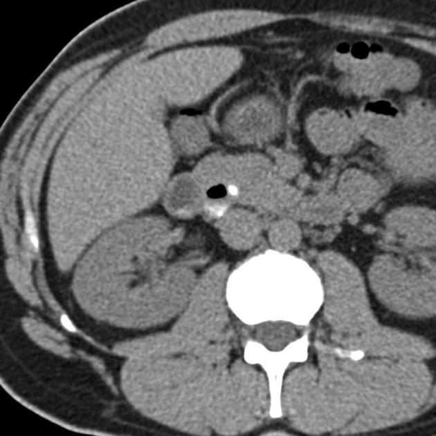
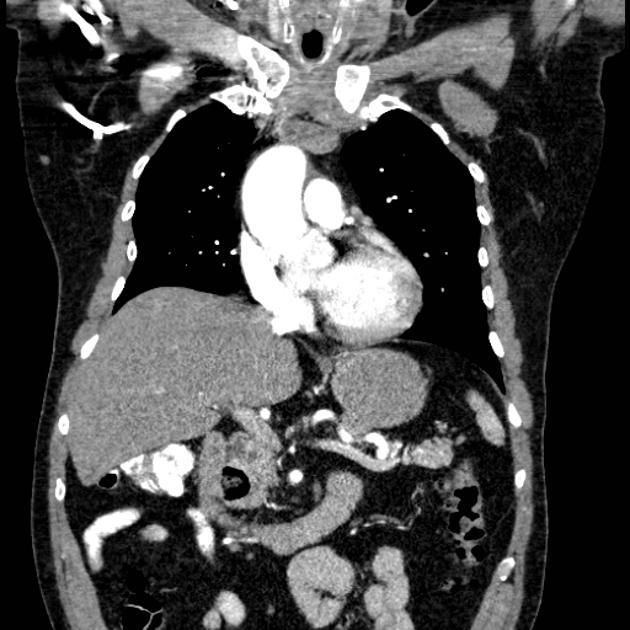
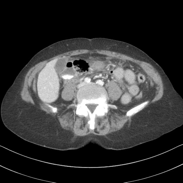
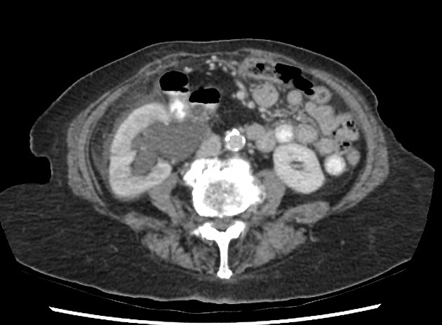
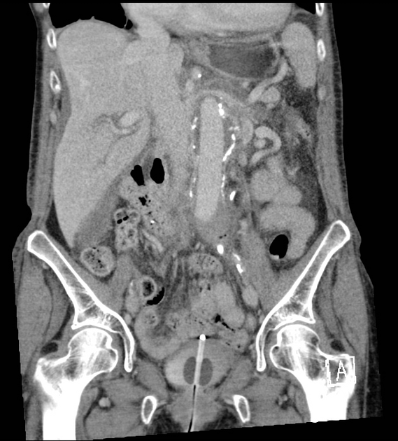
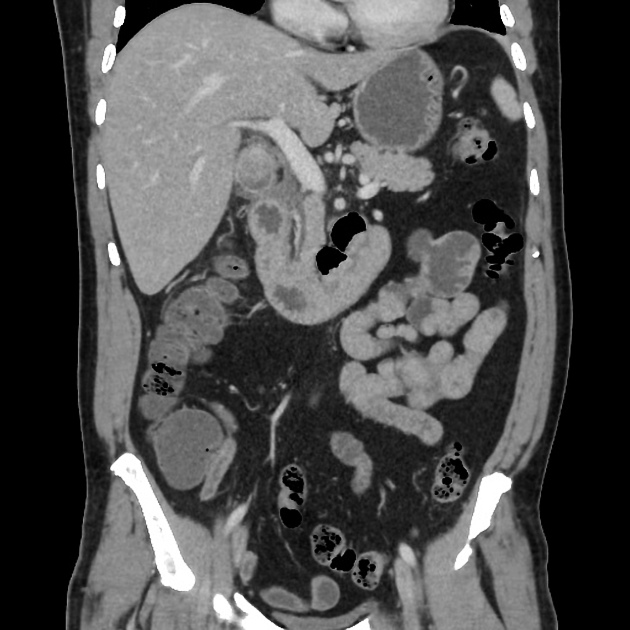
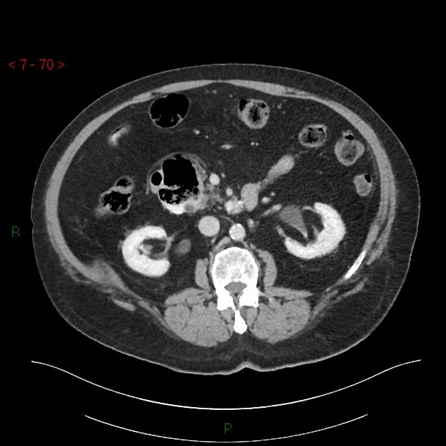
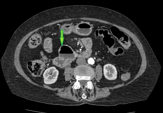
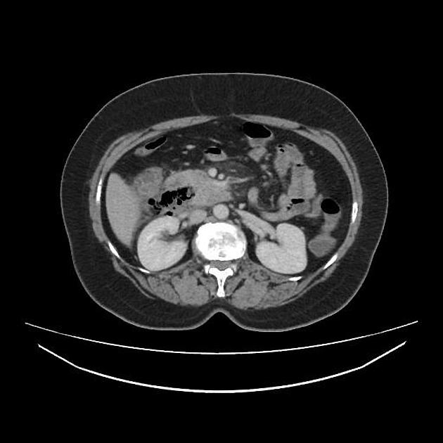
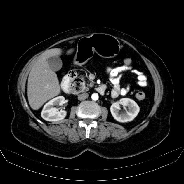
CT
Diverticula are seen as saccular outpouchings from the duodenum that may contain gas, fluid, contrast, or food debris or any combination of these. They often contain a gas-fluid or gas-contrast level 3.
Treatment and prognosis
Complications
Potential complications include 2,3
hemorrhage
perforation
abscess formation
biliary duct obstruction - Lemmel syndrome
Differential diagnosis
The major consideration and mimic of a duodenal diverticulum are that of extraluminal periduodenal gas, which can be caused by:
duodenal injury - iatrogenic or trauma
An extraluminal periduodenal collection of fluid may also be caused by:


 Unable to process the form. Check for errors and try again.